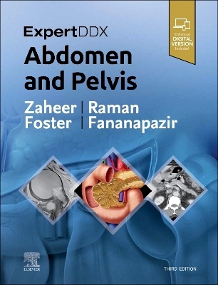
ExpertDDx: Abdomen and Pelvis
Elsevier - Health Sciences Division (Verlag)
978-0-323-87866-1 (ISBN)
Covers 175 of the most common diagnostic challenges in abdominal and pelvic imaging, enhanced by more than 2,100 radiologic images, full-color illustrations, clinical and histologic photographs, and gross pathology images
Provides a quick review of the salient features of each entity, differentiating features from other similar-appearing abnormalities
Includes new chapters on hematuria, flank pain, acute scrotal pain, and seminal vesicle
Adds greater focus to advancing prostate imaging methods with expanded content on lesions in the peripheral zone and lesions in the transition zone, as well as new coverage of transplant imaging
Contains updates to numerous classifications, including LI-RADS for liver, O-RADS for ovarian masses, and the Tanaka classification for pancreatic cysts
Features new MR examples and MR-specific diagnoses throughout, plus new differentials for contrast-enhanced ultrasound findings related to liver and kidney lesions
Includes the enhanced eBook version, which allows you to search all text, figures, and references on a variety of devices
Dr. Atif Zaheer has a specialty focus on CT, MR, and US of the abdomen and pelvis. He is Professor of Radiology, Oncology and Medicine at The Johns Hopkins University School of Medicine in Baltimore, Maryland. Dr. Siva Raman is a board-certified radiologist who gained his subspecialty expertise in thoracoabdominal imaging during a fellowship at Stanford University, and intern residency at UC Davis Medical Center. He attended Johns Hopkins University Medical School. Dr. Raman is a radiologist at Bay Imaging Consultants in Walnut Creek, California, with a subspecialty focus on thoracoabdominal imaging Dr. Bryan Foster is a body imaging radiologist with a subspecialty focus on oncologic, hepatobiliary, pancreatic, prostate, and small bowel imaging. Dr. Foster is the director of ultrasound at OHSU and performs complex image-guided biopsies; he also performs CEUS imaging and US elastography. Dr. Foster is skilled in all facets of imaging including radiography, fluoroscopy, ultrasound, CT, and MRI. His interests include oncologic, hepatobiliary, pancreatic, prostate, and small bowel imaging. Dr. Foster has recently started performing contrast-enhanced ultrasound imaging (as the only site in Oregon) and US elastography. He is one of two radiologists that perform MRI-guided prostate biopsies (also only site in Oregon). Recently he was selected as an Honored Educator for the Radiological Society of North America. Dr. Ghaneh Fananapazir is a radiologist with specialization in abdominal radiology. He is Professor of Radiology at the University of California, Davis, in Sacramento, California. Fananapazir's clinical focus includes nonvascular interventional procedures, advanced US, CT and MR techniques, vascular imaging, and abdominal organ transplantation (kidney, liver, pancreas). In addition to his clinical duties, Dr. Fananapazir is passionate about medical education and research, particularly relating to kidney transplantation, and has authored textbooks and peer-reviewed research in highly impactful medical journals.
SECTION 1: PERITONEUM AND
MESENTERY
GENERIC IMAGING PATTERNS
4 Mesenteric or Omental Mass (Solid)
Siva P. Raman, MD
10 Mesenteric or Omental Mass (Cystic)
Siva P. Raman, MD
14 Fat-Containing Lesion, Peritoneal Cavity
Siva P. Raman, MD
18 Mesenteric Lymphadenopathy
Siva P. Raman, MD
22 Abdominal Calcifications
Siva P. Raman, MD
28 Pneumoperitoneum
Siva P. Raman, MD
32 Hemoperitoneum
Siva P. Raman, MD
36 Misty (Infiltrated) Mesentery
Siva P. Raman, MD
MODALITY-SPECIFIC IMAGING FINDINGS
COMPUTED TOMOGRAPHY
42 High-Attenuation (Hyperdense) Ascites
Siva P. Raman, MD
SECTION 2: ABDOMINAL WALL
ANATOMICALLY BASED DIFFERENTIALS
48 Abdominal Wall Mass
Siva P. Raman, MD
52 Mass in Iliopsoas Compartment
Siva P. Raman, MD
54 Groin Mass
Siva P. Raman, MD
58 Elevated or Deformed Hemidiaphragm
Siva P. Raman, MD
60 Defect in Abdominal Wall (Hernia)
Siva P. Raman, MD
SECTION 3: ESOPHAGUS
GENERIC IMAGING PATTERNS
66 Intraluminal Mass, Esophagus
Atif Zaheer, MD and Michael P. Federle, MD, FACR
68 Extrinsic Mass, Esophagus
Atif Zaheer, MD and Michael P. Federle, MD, FACR
72 Lesion at Pharyngoesophageal Junction
Atif Zaheer, MD and Michael P. Federle, MD, FACR
74 Esophageal Ulceration
Atif Zaheer, MD and Michael P. Federle, MD, FACR
76 Mucosal Nodularity, Esophagus
Atif Zaheer, MD and Michael P. Federle, MD, FACR
78 Esophageal Strictures
Atif Zaheer, MD and Michael P. Federle, MD, FACR
80 Dilated Esophagus
Atif Zaheer, MD and Michael P. Federle, MD, FACR
82 Esophageal Outpouchings (Diverticula)
Atif Zaheer, MD and Michael P. Federle, MD, FACR
84 Esophageal Dysmotility
Atif Zaheer, MD and Michael P. Federle, MD, FACR
CLINICALLY BASED DIFFERENTIALS
86 Odynophagia
Atif Zaheer, MD and Michael P. Federle, MD, FACR
SECTION 4: STOMACH
GENERIC IMAGING PATTERNS
90 Gastric Mass Lesions
Atif Zaheer, MD and Michael P. Federle, MD, FACR
96 Intramural Mass, Stomach
Atif Zaheer, MD and Michael P. Federle, MD, FACR
98 Target or Bull's-Eye Lesions, Stomach
Atif Zaheer, MD and Michael P. Federle, MD, FACR
100 Gastric Ulceration (Without Mass)
Atif Zaheer, MD and Michael P. Federle, MD, FACR
102 Intrathoracic Stomach
Atif Zaheer, MD and Michael P. Federle, MD, FACR
104 Thickened Gastric Folds
Atif Zaheer, MD and Michael P. Federle, MD, FACR
110 Gastric Dilation or Outlet Obstruction
Atif Zaheer, MD and Michael P. Federle, MD, FACR
114 Linitis Plastica, Limited Distensibility
Atif Zaheer, MD and Michael P. Federle, MD, FACR
CLINICALLY BASED DIFFERENTIALS
118 Epigastric Pain
Atif Zaheer, MD and Michael P. Federle, MD, FACR
124 Left Upper Quadrant Mass
Atif Zaheer, MD and Michael P. Federle, MD, FACR
SECTION 5: DUODENUM
GENERIC IMAGING PATTERNS
130 Duodenal Mass
Atif Zaheer, MD and Michael P. Federle, MD, FACR
136 Dilated Duodenum
Atif Zaheer, MD and Michael P. Federle, MD, FACR
138 Thickened Duodenal Folds
Atif Zaheer, MD and Michael P. Federle, MD, FACR
SECTION 6: SMALL INTESTINE
GENERIC IMAGING PATTERNS
142 Multiple Masses or Filling Defects, Small Bowel
Atif Zaheer, MD and Michael P. Federle, MD, FACR
144 Cluster of Dilated Small Bowel
Atif Zaheer, MD and Michael P. Federle, MD, FACR
146 Aneurysmal Dilation of Small Bowel Lumen
Atif Zaheer, MD and Michael P. Federle, MD, FACR
148 Stenosis, Terminal Ileum
Atif Zaheer, MD and Michael P. Federle, MD, FACR
150 Segmental or Diffuse Small Bowel Wall Thickening
Atif Zaheer, MD and Michael P. Federle, MD, FACR
156 Pneumatosis of Small Intestine or Colon
Atif Zaheer, MD and Michael P. Federle, MD, FACR
CLINICALLY BASED DIFFERENTIALS
160 Occult GI Bleeding
Atif Zaheer, MD and Michael P. Federle, MD, FACR
164 Small Bowel Obstruction
Atif Zaheer, MD and Michael P. Federle, MD, FACR
SECTION 7: COLON
GENERIC IMAGING PATTERNS
172 Solitary Colonic Filling Defect
Atif Zaheer, MD and Michael P. Federle, MD, FACR
174 Multiple Colonic Filling Defects
Atif Zaheer, MD and Michael P. Federle, MD, FACR
176 Mass or Inflammation of Ileocecal Area
Atif Zaheer, MD and Michael P. Federle, MD, FACR
182 Colonic Ileus or Dilation
Atif Zaheer, MD and Michael P. Federle, MD, FACR
186 Toxic Megacolon
Atif Zaheer, MD and Michael P. Federle, MD, FACR
188 Rectal or Colonic Fistula
Atif Zaheer, MD and Michael P. Federle, MD, FACR
194 Segmental Colonic Narrowing
Atif Zaheer, MD and Michael P. Federle, MD, FACR
198 Colonic Thumbprinting
Atif Zaheer, MD and Michael P. Federle, MD, FACR
200 Colonic Wall Thickening
Atif Zaheer, MD and Michael P. Federle, MD, FACR
206 Smooth Ahaustral Colon
Atif Zaheer, MD and Michael P. Federle, MD, FACR
CLINICALLY BASED DIFFERENTIALS
208 Acute Right Lower Quadrant Pain
Atif Zaheer, MD and Michael P. Federle, MD, FACR
214 Acute Left Lower Quadrant Pain
Atif Zaheer, MD and Michael P. Federle, MD, FACR
SECTION 8: SPLEEN
GENERIC IMAGING PATTERNS
222 Splenomegaly
Siva P. Raman, MD
226 Multiple Splenic Calcifications
Siva P. Raman, MD
228 Solid Splenic Mass or Masses
Siva P. Raman, MD
230 Cystic Splenic Mass
Siva P. Raman, MD
MODALITY-SPECIFIC IMAGING FINDINGS
COMPUTED TOMOGRAPHY
232 Diffuse Increased Attenuation, Spleen
Siva P. Raman, MD
SECTION 9: LIVER
GENERIC IMAGING PATTERNS
236 Liver Mass With Central or Eccentric Scar
Atif Zaheer, MD and Michael P. Federle, MD, FACR
240 Focal Liver Lesion With Hemorrhage
Atif Zaheer, MD and Michael P. Federle, MD, FACR
244 Liver "Mass" With Capsular Retraction
Atif Zaheer, MD and Michael P. Federle, MD, FACR
246 Fat-Containing Liver Mass
Atif Zaheer, MD and Michael P. Federle, MD, FACR
248 Cystic Hepatic Mass
Atif Zaheer, MD and Michael P. Federle, MD, FACR
252 Focal Hypervascular Liver Lesion
Atif Zaheer, MD and Michael P. Federle, MD, FACR
258 Liver Mass With Mosaic Enhancement
Atif Zaheer, MD
262 Mosaic or Patchy Hepatogram
Atif Zaheer, MD and Michael P. Federle, MD, FACR
266 Hepatic Calcifications
Atif Zaheer, MD and Michael P. Federle, MD, FACR
270 Liver Lesion Containing Gas
Atif Zaheer, MD and Michael P. Federle, MD, FACR
274 Portal Venous Gas
Atif Zaheer, MD and Michael P. Federle, MD, FACR
276 Widened Hepatic Fissures
Atif Zaheer, MD and Michael P. Federle, MD, FACR
278 Dysmorphic Liver With Abnormal Bile Ducts
Atif Zaheer, MD and Michael P. Federle, MD, FACR
282 Focal Hyperperfusion Abnormality (THAD or THID)
Atif Zaheer, MD and Michael P. Federle, MD, FACR
MODALITY-SPECIFIC IMAGING FINDINGS
MAGNETIC RESONANCE IMAGING
288 Multiple Hypodense Liver Lesions
Atif Zaheer, MD and Michael P. Federle, MD, FACR
294 Multiple Hypointense Liver Lesions (T2WI)
Atif Zaheer, MD and Michael P. Federle, MD, FACR
298 Hyperintense Liver Lesions (T1WI)
Atif Zaheer, MD and Michael P. Federle, MD, FACR
304 Liver Lesion With Capsule or Halo on MR
Atif Zaheer, MD and Michael P. Federle, MD, FACR
COMPUTED TOMOGRAPHY
308 Focal Hyperdense Hepatic Mass on Nonenhanced CT
Atif Zaheer, MD and Michael P. Federle, MD, FACR
312 Periportal Lucency or Edema
Atif Zaheer, MD and Michael P. Federle, MD, FACR
318 Widespread Low Attenuation Within Liver
Atif Zaheer, MD and Michael P. Federle, MD, FACR
ULTRASOUND
322 Focal Hepatic Echogenic Lesion ± Acoustic
Shadowing
Atif Zaheer, MD, Gregory E. Antonio, MD, DRANZCR,
FHKCR, and Eric K. H. Liu, PhD, RDMS
328 Hyperechoic Liver, Diffuse
Atif Zaheer, MD, Gregory E. Antonio, MD, DRANZCR,
FHKCR, and Eric K. H. Liu, PhD, RDMS
330 Hepatomegaly
Hee Sun Park, MD, PhD and Aya Kamaya, MD, FSRU, FSAR
334 Diffuse Liver Disease
Hee Sun Park, MD, PhD and Aya Kamaya, MD, FSRU, FSAR
336 Cystic Liver Lesion
Hee Sun Park, MD, PhD and Aya Kamaya, MD, FSRU, FSAR
340 Hypoechoic Liver Mass
Hee Sun Park, MD, PhD and Aya Kamaya, MD, FSRU, FSAR
344 Echogenic Liver Mass
Hee Sun Park, MD, PhD and Aya Kamaya, MD, FSRU, FSAR
348 Target Lesions in Liver
Hee Sun Park, MD, PhD and Aya Kamaya, MD, FSRU, FSAR
350 Multiple Hepatic Masses
Hee Sun Park, MD, PhD and Aya Kamaya, MD, FSRU, FSAR
354 Hepatic Mass With Central Scar
Hee Sun Park, MD, PhD and Aya Kamaya, MD, FSRU, FSAR
356 Periportal Lesion
Hee Sun Park, MD, PhD and Aya Kamaya, MD, FSRU, FSAR
360 Irregular Hepatic Surface
Aya Kamaya, MD, FSRU, FSAR
362 Portal Vein Abnormality
Hee Sun Park, MD, PhD and Aya Kamaya, MD, FSRU, FSAR
SECTION 10: GALLBLADDER
GENERIC IMAGING PATTERNS
366 Distended Gallbladder
Siva P. Raman, MD
368 Gas in Bile Ducts or Gallbladder
Siva P. Raman, MD
372 Focal Gallbladder Wall Thickening
Siva P. Raman, MD
374 Diffuse Gallbladder Wall Thickening
Jade Wong-You-Cheong, MBChB, MRCP, FRCR, FSRU,
FSAR
MODALITY-SPECIFIC IMAGING FINDINGS
COMPUTED TOMOGRAPHY
378 High-Attenuation (Hyperdense) Bile in Gallbladder
Siva P. Raman, MD
ULTRASOUND
380 Hyperechoic Gallbladder Wall
Jade Wong-You-Cheong, MBChB, MRCP, FRCR, FSRU,
FSAR
382 Echogenic Material in Gallbladder
Jade Wong-You-Cheong, MBChB, MRCP, FRCR, FSRU,
FSAR
384 Dilated Gallbladder
Jade Wong-You-Cheong, MBChB, MRCP, FRCR, FSRU,
FSAR
388 Intrahepatic and Extrahepatic Duct Dilatation
L. Nayeli Morimoto, MD and Aya Kamaya, MD, FSRU,
FSAR
CLINICALLY BASED DIFFERENTIALS
390 Right Upper Quadrant Pain
Siva P. Raman, MD
SECTION 11: BILIARY TRACT
GENERIC IMAGING PATTERNS
398 Dilated Common Bile Duct
Siva P. Raman, MD
404 Asymmetric Dilation of Intrahepatic Bile Ducts
Siva P. Raman, MD
408 Biliary Strictures, Multiple
Siva P. Raman, MD
MODALITY-SPECIFIC IMAGING FINDINGS
MAGNETIC RESONANCE IMAGING
412 Hypointense Lesion in Biliary Tree (MRCP)
Siva P. Raman, MD
SECTION 12: PANCREAS
GENERIC IMAGING PATTERNS
416 Hypovascular Pancreatic Mass
Siva P. Raman, MD
422 Hypervascular Pancreatic Mass
Siva P. Raman, MD
426 Cystic Pancreatic Mass
Siva P. Raman, MD
432 Atrophy or Fatty Replacement of Pancreas
Siva P. Raman, MD
434 Dilated Pancreatic Duct
Siva P. Raman, MD
438 Infiltration of Peripancreatic Fat Planes
Siva P. Raman, MD
444 Pancreatic Calcifications
Siva P. Raman, MD
MODALITY-SPECIFIC IMAGING FINDINGS
ULTRASOUND
448 Cystic Pancreatic Lesion
Fauzia Vandermeer, MD
452 Solid Pancreatic Lesion
Fauzia Vandermeer, MD
456 Pancreatic Duct Dilatation
Fauzia Vandermeer, MD
SECTION 13: RETROPERITONEUM
GENERIC IMAGING PATTERNS
460 Retroperitoneal Mass, Cystic
Matthew T. Heller, MD, FSAR
466 Retroperitoneal Mass, Soft Tissue Density
Matthew T. Heller, MD, FSAR
472 Retroperitoneal Mass, Fat Containing
Matthew T. Heller, MD, FSAR
474 Retroperitoneal Hemorrhage
Matthew T. Heller, MD, FSAR
SECTION 14: ADRENAL
GENERIC IMAGING PATTERNS
478 Adrenal Mass
Mitchell Tublin, MD
SECTION 15: KIDNEY
GENERIC IMAGING PATTERNS
486 Solid Renal Mass
Mitchell Tublin, MD
490 Cystic Renal Mass
Mitchell Tublin, MD
494 Bilateral Renal Cysts
| Erscheinungsdatum | 06.12.2022 |
|---|---|
| Reihe/Serie | ExpertDDx |
| Zusatzinfo | <p>Features <b>more than 2,600 images</b>, including radiologic images, full-color illustrations, clinical and histologic photographs, and gross pathology images </p>; Illustrations |
| Verlagsort | Philadelphia |
| Sprache | englisch |
| Maße | 216 x 276 mm |
| Gewicht | 2420 g |
| Themenwelt | Medizinische Fachgebiete ► Radiologie / Bildgebende Verfahren ► Radiologie |
| ISBN-10 | 0-323-87866-0 / 0323878660 |
| ISBN-13 | 978-0-323-87866-1 / 9780323878661 |
| Zustand | Neuware |
| Haben Sie eine Frage zum Produkt? |
aus dem Bereich


