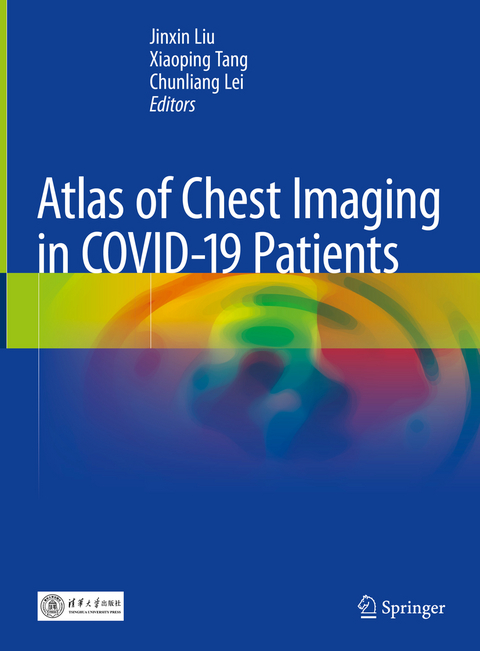
Atlas of Chest Imaging in COVID-19 Patients
Springer Verlag, Singapore
978-981-16-1081-3 (ISBN)
Jinxin Liu is a Professor at Department of Radiology, Guangzhou Eighth People’s Hospital, Guangzhou Medical University, Guangzhou, Guangdong, China Xiaoping Tang is Professor at Department of Infectious Diseases, Guangzhou Medical University, Guangzhou, Guangdong, China Chunliang Lei is a Professor at Department of Infectious Diseases at the same hospital as that of Jinxin Liu.
Overview of COVID-19 Pneumonia.- Common CT Features of COVID-19 Pneumonia.- CT Features of Early COVID-19 Pneumonia (PCR-positive).- CT features of intermediate stage of COVID-19 Pneumonia.- CT Features of late stage of COVID-19 Pneumonia.- Chest Features of Severe and Critical Patients with COVID-19 Pneumonia.- Role of CT and CT features of Suspected COVID-19 Patients (PCR negative).- Follow-up CT of Patients with First Negative CT but Positive PCR for COVID-19.- Imaging Analysis of Family Clustering.- Residual CT Features in Recovery stage of COVID-19 Pneumonia.
| Erscheinungsdatum | 07.05.2021 |
|---|---|
| Zusatzinfo | 6 Illustrations, color; 159 Illustrations, black and white; XII, 192 p. 165 illus., 6 illus. in color. |
| Verlagsort | Singapore |
| Sprache | englisch |
| Maße | 210 x 279 mm |
| Themenwelt | Medizinische Fachgebiete ► Radiologie / Bildgebende Verfahren ► Radiologie |
| Schlagworte | 2019-nCoV • CT images in asymptomatic COVID-19 patients • differential diagnosis of 2019-nCoV • Imaging features of 2019-nCoV • New coronavirus pneumonia |
| ISBN-10 | 981-16-1081-9 / 9811610819 |
| ISBN-13 | 978-981-16-1081-3 / 9789811610813 |
| Zustand | Neuware |
| Informationen gemäß Produktsicherheitsverordnung (GPSR) | |
| Haben Sie eine Frage zum Produkt? |
aus dem Bereich


