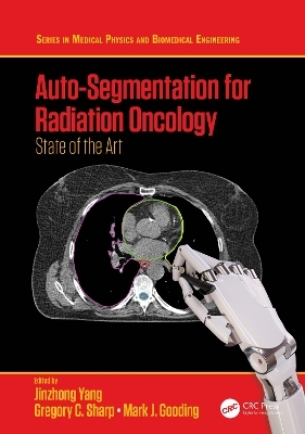
Auto-Segmentation for Radiation Oncology
CRC Press (Verlag)
978-0-367-33600-4 (ISBN)
This book is an ideal guide for radiation oncology centers looking to learn more about potential auto-segmentation tools for their clinic in addition to medical physicists commissioning auto-segmentation for clinical use.
Features:
Up-to-date with the latest technologies in the field
Edited by leading authorities in the area, with chapter contributions from subject area specialists
All approaches presented in this book are validated using a standard benchmark dataset established by the Thoracic Auto-segmentation Challenge held as an event of the 2017 Annual Meeting of American Association of Physicists in Medicine
Jinzhong Yang earned his BS and MS degrees in Electrical Engineering from the University of Science and Technology of China, in 1998 and 2001, and his PhD degree in Electrical Engineering from Lehigh University in 2006. In July 2008, Dr Yang joined the University of Texas MD Anderson Cancer Center as a Senior Computational Scientist, and since January 2015 he has been an Assistant Professor of Radiation Physics. Dr Yang is a board-certified medical physicist. His research interest focuses on deformable image registration and image segmentation for radiation treatment planning and image-guided adaptive radiotherapy, radiomics for radiation treatment outcome modeling and prediction, and novel imaging methodologies and applications in radiotherapy. Greg Sharp earned a PhD in Computer Science and Engineering from the University of Michigan and is currently Associate Professor in Radiation Oncology at Massachusetts General Hospital and Harvard Medical School. His primary research interests are in medical image processing and image-guided radiation therapy, where he is active in the open source software community. Mark Gooding earned his MEng in Engineering Science in 2000 and DPhil in Medical Imaging in 2004, both from the University of Oxford. He was employed as a postdoctoral researcher both in university and hospital settings, where his focus was largely around the use of 3D ultrasound segmentation in women’s health. In 2009, he joined Mirada Medical Ltd, motivated by a desire to see technical innovation translated into clinical practice. While there, he has worked on a broad spectrum of clinical applications, developing algorithms and products for both diagnostic and therapeutic purposes. If given a free choice of research topic, his passion is for improving image segmentation, but in practice he is keen to address any technical challenge. Dr Gooding now leads the research team at Mirada, where in addition to the commercial work he continues to collaborate both clinically and academically.
Contents
Foreword I..........................................................................................................................................ix
Foreword II........................................................................................................................................xi
Editors............................................................................................................................................. xiii
Contributors......................................................................................................................................xv
Chapter 1 Introduction to Auto-Segmentation in Radiation Oncology.........................................1
Jinzhong Yang, Gregory C. Sharp, and Mark J. Gooding
Part I Multi-Atlas for Auto-Segmentation
Chapter 2 Introduction to Multi-Atlas Auto-Segmentation......................................................... 13
Gregory C. Sharp
Chapter 3 Evaluation of Atlas Selection: How Close Are We to Optimal Selection?................. 19
Mark J. Gooding
Chapter 4 Deformable Registration Choices for Multi-Atlas Segmentation............................... 39
Keyur Shah, James Shackleford, Nagarajan Kandasamy, and Gregory C. Sharp
Chapter 5 Evaluation of a Multi-Atlas Segmentation System......................................................49
Raymond Fang, Laurence Court, and Jinzhong Yang
Part II Deep Learning for Auto-Segmentation
Chapter 6 Introduction to Deep Learning-Based Auto-Contouring for Radiotherapy................ 71
Mark J. Gooding
Chapter 7 Deep Learning Architecture Design for Multi-Organ Segmentation......................... 81
Yang Lei, Yabo Fu, Tonghe Wang, Richard L.J. Qiu, Walter J. Curran,
Tian Liu, and Xiaofeng Yang
Chapter 8 Comparison of 2D and 3D U-Nets for Organ Segmentation.................................... 113
Dongdong Gu and Zhong Xue
Chapter 9 Organ-Specific Segmentation Versus Multi-Class Segmentation Using U-Net....... 125
Xue Feng and Quan Chen
Chapter 10 Effect of Loss Functions in Deep Learning-Based Segmentation............................ 133
Evan Porter, David Solis, Payton Bruckmeier, Zaid A. Siddiqui,
Leonid Zamdborg, and Thomas Guerrero
Chapter 11 Data Augmentation for Training Deep Neural Networks ........................................ 151
Zhao Peng, Jieping Zhou, Xi Fang, Pingkun Yan, Hongming Shan, Ge Wang,
X. George Xu, and Xi Pei
Chapter 12 Identifying Possible Scenarios Where a Deep Learning Auto-Segmentation
Model Could Fail...................................................................................................... 165
Carlos E. Cardenas
Part III Clinical Implementation Concerns
Chapter 13 Clinical Commissioning Guidelines......................................................................... 189
Harini Veeraraghavan
Chapter 14 Data Curation Challenges for Artificial Intelligence................................................ 201
Ken Chang, Mishka Gidwani, Jay B. Patel, Matthew D. Li, and
Jayashree Kalpathy-Cramer
Chapter 15 On the Evaluation of Auto-Contouring in Radiotherapy.......................................... 217
Mark J. Gooding
Index............................................................................................................................................... 253
| Erscheinungsdatum | 20.04.2021 |
|---|---|
| Reihe/Serie | Series in Medical Physics and Biomedical Engineering |
| Zusatzinfo | 32 Tables, black and white; 1 Line drawings, color; 56 Line drawings, black and white; 23 Halftones, color; 21 Halftones, black and white; 101 Illustrations, black and white |
| Verlagsort | London |
| Sprache | englisch |
| Maße | 178 x 254 mm |
| Gewicht | 670 g |
| Themenwelt | Medizin / Pharmazie ► Allgemeines / Lexika |
| Medizin / Pharmazie ► Medizinische Fachgebiete ► Onkologie | |
| Medizinische Fachgebiete ► Radiologie / Bildgebende Verfahren ► Radiologie | |
| Naturwissenschaften ► Biologie | |
| Naturwissenschaften ► Physik / Astronomie ► Angewandte Physik | |
| ISBN-10 | 0-367-33600-6 / 0367336006 |
| ISBN-13 | 978-0-367-33600-4 / 9780367336004 |
| Zustand | Neuware |
| Informationen gemäß Produktsicherheitsverordnung (GPSR) | |
| Haben Sie eine Frage zum Produkt? |
aus dem Bereich


