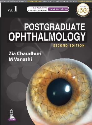
Postgraduate Ophthalmology
Jaypee Brothers Medical Publishers (Verlag)
978-93-89587-33-3 (ISBN)
The new edition of this two-volume set is a complete guide to the eye, and the diagnosis and management of ocular diseases and disorders.
Divided into 18 sections, the book begins with discussion on refraction, retinoscopy and low vision. The next section covers ocular genetics, gene editing, microbiology and pathology, then numerous different imaging techniques.
Each of the following sections provides in depth coverage of different disorders and conditions that may occur in a specific part of the eye. A complete section is dedicated to clinical trials.
The second edition has been fully revised and updated to provide clinicians with the latest advances in the field. Many new topics have also been added to this edition.
The comprehensive text is highly illustrated with nearly 3500 clinical photographs, diagrams and tables across 3300 pages.
Key points
Comprehensive guide to complete field of ophthalmology over two volumes and 3300 pages
Fully revised second edition providing latest advances and featuring many new topics
Highly illustrated with nearly 3500 clinical photographs, diagrams and tables
Previous edition (9789350252703) published in 2011
Zia Chaudhuri MD Professor of Ophthalmology at LHMC & Associated Hospitals, PGIMER & Dr RML Hospital, New Delhi, India M Vanathi MD Associate Professor of Ophthalmology, Dr Rajendra Prasad Centre for Ophthalmic Sciences, All India Institute of Medical Sciences, New Delhi, India
1.1 Light and its Properties
1.2 Reflection, Refraction and Geometric Optics
1.3 Refraction of the Eye: Principles and Practice of Retinoscopy
1.4 Low Vision
1.5 Colour Vision
2.1 Genetics in Ophthalmology
2.2 Gene Editing and Gene Therapy in Inherited Retinal Degeneration
2.3 Ocular Biochemistry
2.4 Ocular Microbiology
2.5 Ocular Pharmacology
2.6 Ocular Pathology
2.7 Anesthesia for Ophthalmic Surgery
2.8 Principles of Ophthalmic Radiology
2.9 Neuroimaging
2.10 Orbital Imaging
2.11 High Resolution Surface Coil Orbital MR Imaging in Strabismus
2.12 Ophthalmic Ultrasonography
2.13 Orbital Ultrasonography
2.14 Anterior Segment OCT
2.15 Posterior Segment OCT - Retina and Macula
2.16 Posterior Segment OCT - Retinal Nerve Fibre Layer
2.17 Adaptive Imaging
2.18 Confocal Microscopy
2.19 Corneal Topography
2.20 Corneal Hysteresis
2.21 Tear Film Imaging
2.22 Psychophysical Tests of the Retina and Macular Function Tests
2.23 Retinal Microperimetry
2.24 Basics of Electrophysiology
2.25 Electrodiagnosis: Basics, Interpretations and Clinical Applications
2.26 Visual Field Evaluation and their Interpretations
2.27 Artificial Intelligence in Ophthalmology
3.1 Cornea: Applied Anatomy and Functions
3.2 Corneal Evaluation
3.3 Bacterial Conjunctivitis and Keratitis
3.4 Viral Conjunctivitis and Keratitis
3.5 Fungal Keratitis
3.6 Acanthamoeba Keratitis
3.7 Microsporidial Keratitis
3.8 Chlamydial Infections of the Eye
3.9 Unusual Corneal Infections: Atypical Mycobacterial Keratitis and Pythium Keratitis
3.10 Corneal Ectasias
3.11 Corneal Dystrophies
3.12 Diseases of the Corneal Limbus & Miscellaneous Conditions of Cornea
3.13 Limbal Stem Cell Deficiency
3.14 Diseases of the Conjunctiva
3.15 Tear Film Dysfunction - Dry Eye Disease
3.16 Immunological Diseases of the Eye
3.17 Diseases of the Sclera
3.18 Contact Lens: Indications and Management
3.19 Penetrating Keratopasty
3.20 Lamellar Keratoplasty
3.21 Ocular Surface Reconstruction with Amniotic Membrane Grafting
3.22 Keratoprosthesis
3.23 Eye Banking
4.1 Laser Refractive Surgery & Phakic IOL
4.2 Lenticule Extraction Refractive Surgery (SMILE)
4.3 Incisional Keratorefractive Surgery
4.4 Corneal Crosslinking
5.1 Glaucoma: Applied Anatomy, Physiology and Classifications
5.2 Glaucoma Evaluation
5.3 Primary Open Angle Glaucoma
5.4 Primary Angle Closure Glaucoma
5.5 Normal Tension Glaucoma
5.6 Secondary Glaucoma
5.7 Medical Management of Glaucoma
5.8 Trabeculectomy and its Modifications
5.9 Glaucoma Drainage Device
5.10 Neuroprotection, Neuro-recovery and Neuro-regeneration
5.11 Hyptony, Atrophic and Phthisis Bulbi
6.1 Lens: Basic Aspects
6.2 Approach to a Patient with Cataract
6.3 Intraocular Lenses
6.4 Intraocular Lens Implant Power Calculations, Selection, Biometry and Pediatric Considerations
6.5 Post-refractive Surgery IOL Power Calculations
6.6 Cataract Surgery
6.7 Small Incision Cataract Surgery
6.8 Femtosecond Laser Assisted Cataract Surgery
6.9 Scleral Fixated Intraocular Lens Implantation
6.10 Management of Posterior Polar Cataract
6.11 Management of Hard Cataract
6.12 Management of Soft Cataract
6.13 Management of White Cataract
6.14 Cataract Surgery in Small Pupils
6.15 Cataract Surgery in Subluxated Cataracts
6.16 Cataract Surgery in Diabetic Patients
6.17 Cataract Surgery in Corneal Disorders
6.18 Cataract Surgery in Eyes with Co-existing Ocular and Systemic Co-morbidity
6.19 Analysis of Surgically Induced Astigmatism
7.1 Anatomy, Physiology and Diseases of the Uvea
7.2 Iridodialysis
7.3 Endophthalmitis
7.4 Panophthalmitis
8.1 Anatomy, Physiology and Diseases of the Vitreous
8.2 Retinal Anatomy
8.3 Retinal Evaluation
8.4 Dystrophy and Degenerations of the Retina
8.5 Retinal Vascular Diseases
8.6 Systemic Diseases with Retinal Manifestations
8.7 Diseases of the Macula
8.8 Retinal Detachment: Etiopathogenesis and Management
9.1 Orbit: Applied Anatomy, Imaging and Evaluation
9.2 Pathological Conditions of the Orbit
9.3 Anophthalmic Socket and Prosthetic Rehabilitation of the Orbit
9.4 Orbital Surgery
9.5 Eyelid Anatomy
9.6 Evaluation and Management of Blepharoptosis
9.7 Eyelid Retraction
9.8 Eyelid Reconstruction
9.9 Eyelid Malpositions
9.10 Nonsurgical Periorbital Esthetic Procedures
9.11 Esthetic Eyelid Surgery
9.12 Botulinum Toxin in Ophthalmic Plastic Surgery
9.13 The Lacrimal System
10.1 Eyelid Tumors
10.2 Orbital Tumors
10.3 Tumors of the Conjunctiva and Ocular Surface
10.4 Intraocular Tumors
11.1 Anatomy of the Visual Pathway
11.2 Neural Basis of Ocular movements
11.3 Pupil
11.4 Cranial Nerve Examination
11.5 Examination Techniques in Neuro-ophthalmology
11.6 Headache and the Eye
11.7 Visual Hallucinations
11.8 Psychosomatic Disorders, Hysteria and Malingering
11.9 Optic Disc Atrophy
11.10 Optic Disc Edema
11.11 Optic Neuritis
11.12 Hereditary Optic Neuropathy
11.13 Ischemic Optic Neuropathy
11.14 Toxic and Nutritional Optic Neuropathy
11.15 Compressive Optic Neuropathy
11.16 Ophthalmoplegia
11.17 Disorders of Supranuclear and Infranuclear Pathways
11.18 Orbit in Neuro-ophthalmic Disorders
11.19 Nystagmus and Other Abnormal Ocular Oscillations
12.1 Visual Functions in Children: Normal Ocular and Visual Development
12.2 Amblyopia
12.3 Pediatric Ocular Examination
12.4 Pediatric Refraction
12.5 Pediatric Refractive Surgery
12.6 Ocular Embryology: Overview and Basis for Congenital Ocular Disorders
12.7 Intrauterine Infections and the Developing Eye
12.8 Congenital and Structural Lesions of the Orbit
12.9 Congenital Eyelid Anomalies
12.10 Diseases of the Pediatric Cornea
12.11 Congenital and Developmental Glaucomas
12.12 Pediatric Lenticular Abnormalities
12.13 Retinopathy of Prematurity
12.14 Recent Advances in Retinopathy of Prematurity
12.15 Child Abuse, Non-accidental Injuries and the Eye
12.16 Craniofacial Malformations
12.17 Ocular Clues to Pediatric Neuro-ophthalmic Diagnosis
12.18 Congenital Optic Disk Anomalies
12.19 Cortical Visual Impairment
13.1 Functional Anatomy and Physiology of Extraocular Muscles
13.2 Physiological Basis of Ocular Misalignment
13.3 Classification of Strabismus
13.4 Clinical Evaluation of Strabismus
13.5 Orthoptic Evaluation and Uses of Orthoptic Instruments
13.6 Primary Comitant Strabismus
13.7 Dissociated Strabismus Complex
13.8 Paralytic Strabismus
13.9 Pattern Strabismus
13.10 Neuroanatomical Strabismus and Pulleopathies
13.11 Strabismus Associated with Neuro-developmental Disorders
13.12 Congenital Cranial Dysinnervation Syndrome
13.13 Duane's Syndrome
13.14 Brown's Syndrome
13.15 MED
13.16 Strabismus in Thyroid Eye Disease
13.17 Post-surgical Strabismus
13.18 Management of Strabismus including Vessel Sparing Procedures
14.1 The Eye in Metabolic and Systemic Disorders
14.2 Albinism and Role of Pigment in Visual Development
14.3 Ophthalmic Manifestations of Leukemia
14.4 The Eye and Carotid Artery Disease
14.5 Ocular Myopathy
14.6 The Eye in Phakomatosis
14.7 Demyelinating Disease and the Eye
14.8 The Eye in Autism Spectrum Disorders
14.9 Visual Manifestations of Stroke
15.1 Ocular Trauma
15.2 Chemical Injuries
15.3 Management of Corneal and Corneo-scleral Tear
15.4 Traumatic Glaucoma & Hyphema
15.5 Traumatic Cataract
15.6 Posterior Segment Ocular Trauma
15.7 Management of Intraocular Foreign Body [IOFB]
15.8 Management of Trauma & Fractures of Orbit
15.9 TON and ONA
15.10 Traumatic Strabismus and Diplopia
15.11 Ocular Manifestations Management of Intracranial Trauma
16.1 Excimer Laser Applications in Cornea
16.2 Femtosecond Laser Applications in Cornea
16.3 Laser Applications in Glaucoma
16.4 YAG Capsulotomy
16.5 Lasers in the Posterior Segment of the Eye
17.1 Cornea Trials
17.2 Glaucoma Trials
17.3 PEDIG and Myopia Trials
17.4 Retina Trials
18.1 Community Ophthalmology
18.2 Ethical and Medicolegal Aspects in Ophthalmology
18.3 Patient Safety, Quality of Care and Accreditation
18.4 Health and Medical Administration: Basic Aspects for an Ophthalmologist
18.5 HAI
18.6 Ophthalmic Emergencies
18.7 Differential Diagnosis of Ophthalmic Signs and Symptoms
19.1 Illustrated Ocular Pathology
19.2 Illustrated Ocular Microbiology
19.3 Radiological Landmarks on Orbital CT Scan
19.4 Neurological Plates
| Erscheinungsdatum | 10.05.2021 |
|---|---|
| Zusatzinfo | 2594 Halftones, color; 840 Illustrations |
| Verlagsort | New Delhi |
| Sprache | englisch |
| Maße | 216 x 279 mm |
| Themenwelt | Medizin / Pharmazie ► Medizinische Fachgebiete ► Augenheilkunde |
| ISBN-10 | 93-89587-33-6 / 9389587336 |
| ISBN-13 | 978-93-89587-33-3 / 9789389587333 |
| Zustand | Neuware |
| Haben Sie eine Frage zum Produkt? |
aus dem Bereich


