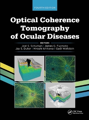
Optical Coherence Tomography of Ocular Diseases
SLACK Incorporated (Verlag)
978-1-63091-708-1 (ISBN)
Written by the pioneers of OCT technologies and the world-renowned OCT researchers Drs. Joel S. Schuman, James G. Fujimoto, Jay S. Duker, Hiroshi Ishikawa, and Gadi Wollstein, Optical Coherence Tomography of Ocular Diseases, Fourth Edition is an essential text for imaging technology.
OCT now occupies a dominant role as a diagnostic tool for retinal conditions and glaucoma. At the same time, the technology continues to show potential for emerging clinical and research applications across all the ophthalmological subspecialties. To reflect these rapid advances, this new edition of Optical Coherence Tomography of Ocular Diseases features a complete and thorough revision of the existing text as well as the addition of cutting-edge content to bring this classic resource completely up to date.
New content in the Fourth Edition includes:
• OCT angiography
• Swept-source OCT
• OCT in multimodal imaging
• Clinical utility of OCT in glaucoma prediction and progression detection
• OCT for neuro-ophthalmology
Optical Coherence Tomography of Ocular Diseases, Fourth Edition is the one and only book needed by practitioners who use OCT for clinical eye care.
Joel S. Schuman, MD, is the Elaine Langone Professor and Vice Chair for Research in the Department of Ophthalmology and Professor of Neuroscience & Physiology at NYU Langone Health, NYU Grossman School of Medicine. He is Professor of Biomedical Engineering and Electrical & Computer Engineering at NYU Tandon School of Engineering and Professor of Neural Science in the Center for Neural Science at NYU College of Arts and Sciences. He chaired the ophthalmology department at NYU Langone Health, NYU Grossman School of Medicine from 2016-2020. Prior to arriving at NYU in 2016, he was the Eye and Ear Foundation Professor and Chairman of Ophthalmology (2003-2016), the Eye and Ear Institute, University of Pittsburgh School of Medicine and Director of the University of Pittsburgh Medical Center Eye Center, Professor of Bioengineering at the Swanson School of Engineering, University of Pittsburgh and Founder of the Louis J. Fox Center for Vision Restoration of the University of Pittsburgh Medical Center and the University of Pittsburgh. He was a member of the McGowan Institute for Regenerative Medicine and the Center for the Neural Basis of Cognition, Carnegie Mellon University, and University of Pittsburgh. Dr. Schuman is a native of Roslyn, New York; he graduated Columbia University (AB, 1980) and Mt. Sinai School of Medicine (MD, 1984). Following his internship at New York’s Beth Israel Medical Center (1985), he completed residency training at the Medical College of Virginia (1988) and a glaucoma fellowship at Massachusetts Eye & Ear Infirmary (clinical 1989; research 1990), where he was a Heed Fellow. After just over 1 year on the Harvard faculty, he moved to New England Medical Center, Tufts University to cofound the New England Eye Center in 1991, where he was Residency Director (1991-1999) and Glaucoma and Cataract Service Chief (1991-2003). He became a Professor of Ophthalmology in 1998 and Vice Chair in 2001. Dr. Schuman and his colleagues were first to identify a molecular marker for human glaucoma, published in Nature Medicine in 2001. Continuously funded by the National Eye Institute as a principal investigator since 1995, he is an inventor of optical coherence tomography (OCT), used world-wide for ocular diagnostics. Dr. Schuman has published more than 400 peer-reviewed scientific journal articles, has authored or edited 8 books, and has contributed more than 80 book chapters. Dr. Schuman is a founding member of the Association for Research in Vision and Ophthalmology (ARVO) Multidisciplinary Ophthalmic Imaging (MOI) cross-sectional group, served on the program committee from its founding and chaired the MOI program committee from 2007-2013. He is also a founder and cochair of ARVO Imaging (formerly ARVO/ISIE, The International Society for Imaging in the Eye, inaugurated 2002). Dr. Schuman was cochair of the International Glaucoma Symposium from 1998-2007, the world’s largest meeting devoted to glaucoma, which merged with the World Glaucoma Congress in 2007, for which he was Program cochair from 2007-2011. He chaired the Hawaiian Eye meeting glaucoma section from 1993-2019. In 2002, he received the Alcon Research Institute Award and the Lewis Rudin Glaucoma Prize; in 2003, the Senior Achievement Award from the American Academy of Ophthalmology (AAO); in 2004, he was elected into the American Society for Clinical Investigation; in 2006, he received the ARVO Translational Research Award; and in 2012, the Carnegie Science Center Award as well as sharing the Champalimaud Award (a 1 million Euro cash prize). He was elected to the American Ophthalmological Society in 2008. In 2011, Dr. Schuman was the Clinician-Scientist Lecturer of the American Glaucoma Society. In 2012, he received the Carnegie Science Center’s Award in Life Sciences. In 2013, he gave the Robert N. Shaffer Lecture at the American AAO Annual Meeting and received the AAO Lifetime Achievement Award. In 2014, he became a Gold Fellow of ARVO, and he received a Special Recognition Award from the AAO in 2015. He was elected to the American Association of Physicians and also received the Fight for Sight Physician/Scientist Award in 2016. In 2017, he received the Leslie Dana Gold Medal. In 2018, Dr. Schuman was the American Glaucoma Society Lecturer and received the Fight for Sight Alumni Achievement Award. In 2019, he was given the BrightFocus Scientific Impact Award, and in 2021, the Eye Foundation of America Robert Murphy Visionary Award. He is named in Who’s Who in America, Who’s Who in Medical Sciences Education, America’s Top Doctors, and Best Doctors in America. James G. Fujimoto, PhD, is Elihu Thomson Professor of Electrical Engineering at the Massachusetts Institute of Technology, visiting Professor of Ophthalmology at Tufts University School of Medicine, and Adjunct Professor at the Medical University of Vienna. His group and collaborators were responsible for the invention and development of OCT in the early 1990s. Dr. Fujimoto cofounded the startup company Advanced Ophthalmic Devices, which developed ophthalmic OCT imaging, and was acquired by Carl Zeiss and also cofounded LightLab Imaging, which developed cardiovascular OCT, and was acquired by Goodman, Ltd. He received the 2011 Zeiss Research Award and the 2015 Optical Society of America Ives Medal and is a corecipient of the 2002 Rank Prize in Optoelectronics, the 2012 Antonio Champalimaud Vision Award, the 2017 European Inventor Award, the 2017 National Academy of Engineering Fritz J. and Dolores H. Russ Prize, and the Greenberg End Blindness Award 2020. Dr. Fujimoto is a member of the National Academy of Engineering, National Academy of Sciences, and American Academy of Arts and Sciences, and he has honorary doctorates from the Friedrich-Alexander-Universität Erlangen-Nürnberg, Germany and Nicolaus Copernicus University in Torun, Poland. Jay S. Duker, MD, is the Ophthalmologist-in-Chief at Tufts Medical Center, Director of the New England Eye Center, and Chairman of the Department of Ophthalmology at Tufts University School of Medicine. Dr. Duker has been at Tufts Medical Center for more than 30 years and previously served as Director of the Retina Service. A graduate of Harvard University and Jefferson Medical College, he completed his postgraduate training at Wills Eye Hospital in Philadelphia, Pennsylvania. His research interests include new treatments of vascular disease, intraocular drug delivery, and imaging of the posterior segment. He has been instrumental in the development of OCT. Hiroshi Ishikawa, MD, completed medical school in 1989 at Mie University School of Medicine (Mie, Japan). After completion of a glaucoma fellowship in Japan, he became a glaucoma research fellow under Dr. Robert Ritch at New York Eye & Ear Infirmary in 1996. He is currently a Professor of Ophthalmology at Oregon Health & Science University (Portland, Oregon). His research revolves around ocular imaging and processing. His research has yielded 19 intellectual properties, 9 US patents allowed, 2 US patents pending, and 8 copy rights. He is recognized as the first inventor of the detailed macular retinal layer segmentation of OCT images. Recently, his main research interest has shifted to machine learning and artificial intelligence in ophthalmology. Gadi Wollstein, MD, is a Professor of Ophthalmology, Bioengineering, and Neural Science and the Director of the Advanced Ophthalmic Imaging Laboratory in NYU, New York, New York. After completing his clinical training in Israel, Dr. Wollstein has been a research fellow in ocular imaging in Moorfields Eye Hospital in London, United Kingdom followed by a second fellowship in Tufts-New England Medical Center, Boston, Massachusetts, where he investigated the use of imaging devices to detect and follows patients with glaucoma. Before joining NYU, Dr. Wollstein directed the lab in the University of Pittsburgh, Pittsburgh, Pennsylvania, for more than a decade. Dr. Wollstein’s research primarily focuses on the use of ocular imaging devices to detect, monitor, and understand the pathophysiology of glaucoma. His studies were summarized in more than 200 peer review manuscripts and more than 50 book chapters.
Dedication About the Editors About the Assistant Editor Contributing AuthorsPreface Section I Principles of Operation and Interpretation Chapter 1 Introduction to Optical Coherence Tomography James G. Fujimoto, PhD; Eric M. Moult, BEng; David Huang, MD, PhD; Joel S. Schuman, MD;Jay S. Duker, MD; Carmen A. Puliafito, MD, MBA; and Eric Swanson, MSIntroductionThe Early Development of Optical Coherence TomographyOptical Coherence TomographyHigh-Speed Spectral/Fourier-Domain Optical Coherence TomographyEn Face Optical Coherence Tomography ImagingUltrahigh-Speed Swept-Source/Fourier-Domain Optical Coherence TomographyOptical Coherence Tomography AngiographyConclusion Chapter 2 Interpretation of the Optical Coherence Tomography Image Hiroshi Ishikawa, MD; Gadi Wollstein, MD; Katie A. Lucy, BS;Lindsey S. Folio, MS, MBA; Jessica E. Nevins, MS; James G. Fujimoto, PhD;Joel S. Schuman, MD; and Carmen A. Puliafito, MD, MBAIntroductionInterpreting Optical Coherence Tomography Images of the Normal RetinaInterpreting Optical Coherence Tomography Images of the Normal Anterior EyeOptical Coherence Tomography Scanning and Imaging ProtocolsQuantitative Measurements of Retinal MorphologyQuality, Artifacts, and Errors in Optical Coherence Tomography ImagesConclusion Section II Optical Coherence Tomography in Retinal Diseases Chapter 3 Vitreoretinal Interface Disorders Shilpa J. Desai, MD; Heeral R. Shah, MD; Elias C. Mavrofrides, MD;Adam H. Rogers, MD; Steven N. Truong, MD; Carmen A. Puliafito, MD, MBA;James G. Fujimoto, PhD; and Jay S. Duker, MDIdiopathic Epiretinal MembraneVitreomacular Traction SyndromeIdiopathic Macular Hole Chapter 4 Retinal Vascular Diseases Vanessa Cruz-Villegas, MD; Heeral R. Shah, MD; James G. Fujimoto, PhD; Jay S. Duker, MD;Carmen A. Puliafito, MD, MBA; and Shilpa J. Desai, MDBranch Retinal Artery OcclusionCentral Retinal Artery OcclusionBranch Retinal Vein OcclusionCentral Retinal Vein OcclusionBilateral Idiopathic Juxtafoveal Retinal TelangiectasisRetinal Arterial Macroaneurysm Chapter 5 Diabetic Retinopathy James Lin, MD; Jin Yang, MD, PhD; Harry W. Flynn Jr, MD Chapter 6 Central Serous Chorioretinopathy Shilpa J. Desai, MD; Heeral R. Shah, MD; Elias C. Mavrofrides, MD;Carmen A. Puliafito, MD, MBA; James G. Fujimoto, PhD; and Jay S. Duker, MDTypical Central Serous ChorioretinopathyChronic Central Serous ChorioretinopathyBullous Central Serous ChorioretinopathyLaser Treatment for Central Serous ChorioretinopathyCentral Serous Chorioretinopathy RTVue-100Photodynamic Therapy Treatment for Central Serous Chorioretinopathy Chapter 7 Age-Related Macular Degeneration Shilpa J. Desai, MD; Heeral R. Shah, MD; Elias C. Mavrofrides, MD; Natalia Villate, MD;Carmen A. Puliafito, MD, MBA; and Jay S. Duker, MDDrusenAdult Vitelliform DystrophyGeographic AtrophyOccult Choroidal NeovascularizationVascularized Pigment Epithelial DetachmentRetinal Angiomatous ProliferationRetinal Pigment Epithelium TearSubretinal HemorrhageDisciform Scar Chapter 8 Pathologic Myopia Heeral R. Shah, MD and Shilpa J. Desai, MDPathologic MyopiaAngioid StreaksMyopic Choroidal NeovascularizationMyopic Macular HoleMyopic SchisisRhegmatogenous Retinal Detachment Chapter 9 Chorioretinal Inflammatory and Infectious Diseases Shilpa J. Desai, MD; Heeral R. Shah, MD; Natalia Villate, MD; Elias C. Mavrofrides, MD;Janet Davis, MD; Jay S. Duker, MD; and Carmen A. Puliafito, MD, MBA Chapter 10 Retinal Dystrophies Vanessa Cruz-Villegas, MD; Heeral R. Shah, MD; Jay S. Duker, MD;Shilpa J. Desai, MD; and Carmen A. Puliafito, MD, MBARetinitis PigmentosaStargardt DiseaseBest DiseaseAdult-Onset Foveomacular Vitelliform Dystrophy Chapter 11 Miscellaneous Retinal Diseases Shilpa J. Desai, MD; Heeral R. Shah, MD; Elias C. Mavrofrides, MD; Vanessa Cruz-Villegas, MD; Carmen A. Puliafito, MD, MBA; and Jay S. Duker, MDCystoid Macular EdemaPosterior Segment TraumaOptic Nerve Pit Section III Optical Coherence Tomography in Glaucoma,Neuro-Ophthalmology, and the Anterior Segment Chapter 12 Clinical Utility of Optical Coherence Tomography in Glaucoma Gadi Wollstein, MD; Maria de los Angeles Ramos Cadena, MD; Katie A. Lucy, BS; Lindsey S. Folio, MS, MBA; Jessica E. Nevins, MS; and Joel S. Schuman, MDNormalPreperimetric/Early GlaucomaModerate GlaucomaAdvanced GlaucomaUtility of Imaging in Longitudinal Glaucoma Assessment Chapter 13 Optical Coherence Tomography for Neuro-Ophthalmology Rithambara Ramachandran, MD; Carlos Mendoza-Santiesteban, MD;Matthew D. Lazzara, MD; Thomas R. Hedges III, MD; Steven Galetta, MD;Laura Balcer, MD, MSCE; and Rachel Nolan-Kenney, MPhilProving Maculopathy That May Mimic Optic NeuropathyOptic Neuropathy in the Presence of RetinopathyFunctional Loss of VisionOptic Nerve SwellingCompressive Optic NeuropathyToxic, Hereditary, and Nutritional Optic NeuropathyOptic NeuritisMultiple Sclerosis, Neuromyelitis Optica, and Anti–Myelin OligodendrocyteGlycoprotein SyndromesMicrocystic Macular EdemaNeurodegenerative DiseasesOptical Coherence Tomography AngiographyConclusion Chapter 14 Anterior Segment Optical Coherence Tomography Yuli Yang, MD, PhD; Yan Li, PhD; and David Huang, MD, PhDIntroductionOptical Coherence Tomography DevicesInterpretation of Corneal ImagesAngle AssessmentRefractive SurgerySmall Incision Lenticule ExtractionCorneal Pathologies and Surgeries Section IV AppendixAppendix Optical Coherence Tomography Scanning and Image-Processing Protocols Hiroshi Ishikawa, MD; Gadi Wollstein, MD; Lindsey S. Folio, MS, MBA;Jessica E. Nevins, MS; James G. Fujimoto, PhD; and Joel S. Schuman, MDLine and Radial Scan ProtocolsCircle Scan ProtocolsCompound Radial and Concentric Circles Scan ProtocolsThree-Dimensional Cube Raster Scan ProtocolsEye Motion TrackingImage-Processing ProtocolsRepeated Scan (Image) AveragingSegmentationC-ModeCircumpapillary Retinal Thickness and Retinal Layer Thickness MapOptic Nerve HeadMaculaProgression AnalysisAnterior Segment Optical Coherence Tomography Scan ProtocolsFinancial Disclosures Index
| Erscheinungsdatum | 10.08.2020 |
|---|---|
| Verlagsort | Thorofare |
| Sprache | englisch |
| Maße | 216 x 279 mm |
| Gewicht | 2177 g |
| Themenwelt | Medizin / Pharmazie ► Medizinische Fachgebiete ► Augenheilkunde |
| ISBN-10 | 1-63091-708-7 / 1630917087 |
| ISBN-13 | 978-1-63091-708-1 / 9781630917081 |
| Zustand | Neuware |
| Haben Sie eine Frage zum Produkt? |
aus dem Bereich


