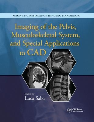
Imaging of the Pelvis, Musculoskeletal System, and Special Applications to CAD
CRC Press (Verlag)
978-0-367-86890-1 (ISBN)
Magnetic resonance imaging (MRI) is a technique used in biomedical imaging and radiology to visualize internal structures of the body. Because MRI provides excellent contrast between different soft tissues, the technique is especially useful for diagnostic imaging of the brain, muscles, and heart.
In the past 20 years, MRI technology has improved significantly with the introduction of systems up to 7 Tesla (7 T) and with the development of numerous post-processing algorithms such as diffusion tensor imaging (DTI), functional MRI (fMRI), and spectroscopic imaging. From these developments, the diagnostic potentialities of MRI have improved impressively with an exceptional spatial resolution and the possibility of analyzing the morphology and function of several kinds of pathology.
Given these exciting developments, the Magnetic Resonance Imaging Handbook: Imaging of the Pelvis, Musculoskeletal System, and Special Applications to CAD is a timely addition to the growing body of literature in the field. Offering comprehensive coverage of cutting-edge imaging modalities, this book:
Discusses MRI of the urinary system, pelvis, spine, soft tissues, lymphatics, and brain
Explains how MRI can be used in fetal, pediatric, forensic, postmortem, and computer-aided diagnostic (CAD) applications
Highlights each organ’s anatomy and pathological processes with high-quality images
Examines the protocols and potentialities of advanced MRI scanners such as 7 T systems
Includes extensive references at the end of each chapter to enhance further study
Thus, the Magnetic Resonance Imaging Handbook: Imaging of the Pelvis, Musculoskeletal System, and Special Applications to CAD provides radiologists and imaging specialists with a valuable, state-of-the-art reference on MRI.
Luca Saba is full professor of Radiology in the University of Cagliari, Italy. Prof. Saba currently works at the Azienda Ospedaliero Universitaria of Cagliari. During his career, he has won 15 scientific and extracurricular awards, published more than 220 papers in high impact factor journals, presented more than 500 papers and posters at national and international congress events, spoken more than 45 times at national and international conferences, written 21 book chapters, edited 14 books, and reviewed more than 40 scientific journals. He is a member of the Italian Society of Medical Radiology, European Society of Radiology, Radiological Society of North America, American Roentgen Ray Society, and European Society of Neuroradiology.
Magnetic Resonance Imaging of Kidneys and Ureters. Carcinoma of the Bladder and Urethra. Male Pelvis (Prostate, Seminal Vesicles, and Testes). Uterus and Vagina. Benign and Malignant Conditions of the Ovaries and Peritoneum. MRI of the Placenta and the Pregnant Patient. MRI of Pelvic Floor Disorders. Degenerative Disease of the Spine and Other Spondyloarthropathies. Spine Infections. Traumatic Disease of the Spine. Neoplastic Disease of the Spine. MR Pathology of Sacrum and Ilium. Magnetic Resonance Imaging of Soft Tissues. Temporomandibular Joints. MR-Guided Interventional Radiology of the Musculoskeletal System. MR of the Lymphatics. Pediatric Applications. Fetal MRI. Postmortem and Forensic Magnetic Resonance Imaging. Magnetic Resonance-Guided Focused Ultrasound. PET/MRI: Concepts and Clinical Applications. Computer-Aided Diagnosis with MR Images of the Brain.
| Erscheinungsdatum | 23.12.2019 |
|---|---|
| Verlagsort | London |
| Sprache | englisch |
| Maße | 210 x 280 mm |
| Gewicht | 1061 g |
| Themenwelt | Medizin / Pharmazie ► Medizinische Fachgebiete ► Orthopädie |
| Medizin / Pharmazie ► Medizinische Fachgebiete ► Radiologie / Bildgebende Verfahren | |
| ISBN-10 | 0-367-86890-3 / 0367868903 |
| ISBN-13 | 978-0-367-86890-1 / 9780367868901 |
| Zustand | Neuware |
| Haben Sie eine Frage zum Produkt? |
aus dem Bereich


