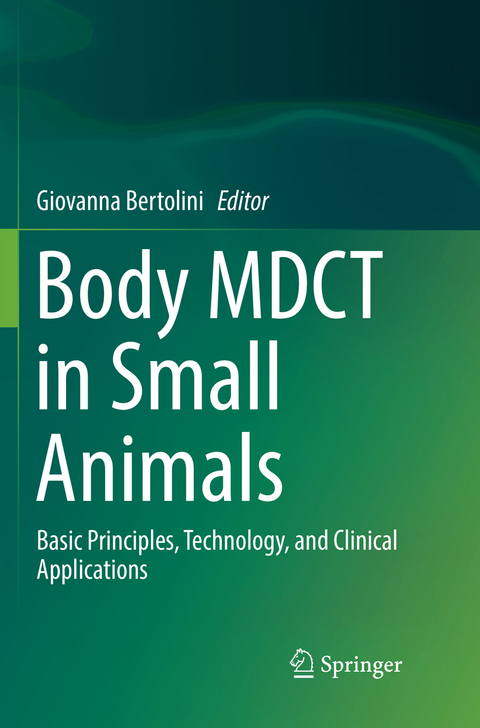
Body MDCT in Small Animals
Springer International Publishing (Verlag)
978-3-319-83617-1 (ISBN)
This book is an up-to-date, technically detailed yet easy-to-read reference book on current clinical applications of MDCT in small animals. It has been designed to serve as the reference book for all MDCT-users, such as veterinary radiologists, imaging technicians, oncologists, surgeons, and non-radiologist clinicians. Individual chapters on novel clinically important topics include applications in endocrinology, oncology, trauma, and cardiovascular CT, as well as sections on organ-specific pathologies and their CT characteristics. The book will also cover main domains of CT, such as thorax and the trauma imaging. Anatomy, clinical aspects, pathology, and CT signs are integrated to provide the reader with the basis for interpretation of MDCT findings. Many excellent 2D multiplanar and 3D figures illustrating typical CT findings of various conditions will serve as a clinical reference for the reader.
Dr. Giovanna Bertolini, DVM, Ph.D, is Head of Diagnostic and Interventional Radiology Division of the San Marco Veterinary Clinic of Padova, Italy since 2003. As one of the first users of MDCT in pets in the world, she developed scanning techniques and protocols for most of the applications in veterinary practice and collected a wide caseload. In the past decade, she concentrated her research on body MDCT in dogs and cats, particularly on vascular and perfusion imaging. Dr. Bertolini received her DVM from the University of Bologna and her Ph.D with special mention of Doctor Europaeus in Veterinary and Comparative Biomedical Sciences at the University of Padova. In 2008 she obtained a Ph.D-research fellowship at the University Medical Centre of Utrecht were she joined the research group of Prof. Mathias Prokop, MD, to investigate post-processing techniques for deriving high-quality CT angiographic data from cerebral MDCT perfusion data. From 2009 to 2013, she was Contract Professor of the `Master of Diagnostic Imaging of the dogs and cats' at the University of Camerino, Italy. She is author of a book `MDCT-row computed tomography for abdominal vascular assessment in dogs' - Lambert Academic Publishing 2010, and author of the Chapter `Tomografia Computerizzata' in the book `Malattie Respiratorie del cane e del gatto' edited by Elsevier 2012. She (co-)authored 28 articles and over 30 abstracts on MDCT in small animals published in international peer-reviewed journals. Her current research interest focuses on various clinical applications of dual-energy CT in small animals.
1 MDCT BASIC PRINCIPLES.- 2 MDCT OF THE THORAX.- MDCT thoracic anatomy.- Larynx, trachea and bronchi.- The lung patterns.- The mediastinum (excluding the heart) and pleura.- The thoracic wall.- 3 MDCT OF THE ABDOMEN.- MDCT anatomy of the abdomen.- The liver.- The pancreas.- The gastrointestinal tract.- The urinary tract.- The peritoneum and abdominal wall.- nodes and small glands.- 4 MDCT OF VASCULAR ANOMALIES.- MDCT vascular anatomy.- Congenital and acquired anomalies of the arterial system.- Congenital and acquired anomalies of the venous system.- Congenital and acquired anomalies of the portal system.
"Intended primarily for radiologists and radiologists in training, the book also could benefit imaging technicians, as it covers imaging protocols with detailed information on angiographic studies. ... This book brings an innovative and needed approach to reviewing computed tomography in veterinary medicine and is written in a clear and organized manner. The very large number of high-quality images as well as the detailed information on angiographic studies and vascular diseases make this book unique." (Cintia R. Oliveira, Doody's Book Reviews, January, 2018)
“Intended primarily for radiologists and radiologists in training, the book also could benefit imaging technicians, as it covers imaging protocols with detailed information on angiographic studies. … This book brings an innovative and needed approach to reviewing computed tomography in veterinary medicine and is written in a clear and organized manner. The very large number of high‐quality images as well as the detailed information on angiographic studies and vascular diseases make this book unique.” (Cintia R. Oliveira, Doody's Book Reviews, January, 2018)
| Erscheint lt. Verlag | 14.8.2018 |
|---|---|
| Zusatzinfo | XV, 453 p. 496 illus., 178 illus. in color. |
| Verlagsort | Cham |
| Sprache | englisch |
| Maße | 155 x 235 mm |
| Gewicht | 964 g |
| Themenwelt | Medizinische Fachgebiete ► Radiologie / Bildgebende Verfahren ► Radiologie |
| Naturwissenschaften ► Biologie ► Zoologie | |
| Veterinärmedizin | |
| Schlagworte | angiography in small animals • body MDCT in cats and dogs • Computed tomography • diagnostic imaging • diagnostic radiology • MDCT technical principles and handling • veterinary diagnostic imaging scanner |
| ISBN-10 | 3-319-83617-X / 331983617X |
| ISBN-13 | 978-3-319-83617-1 / 9783319836171 |
| Zustand | Neuware |
| Informationen gemäß Produktsicherheitsverordnung (GPSR) | |
| Haben Sie eine Frage zum Produkt? |
aus dem Bereich


