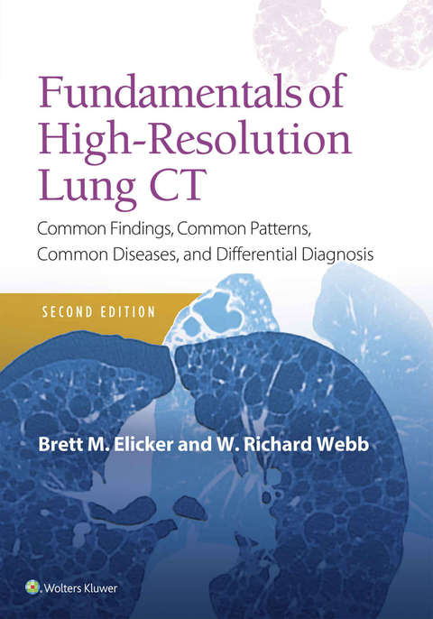
Fundamentals of High-Resolution Lung CT
Common Findings, Common Patterns, Common Diseases and Differential Diagnosis
Seiten
2018
|
2nd edition
Lippincott Williams and Wilkins (Verlag)
978-1-4963-8992-3 (ISBN)
Lippincott Williams and Wilkins (Verlag)
978-1-4963-8992-3 (ISBN)
In clear and concise language, Fundamentals of High-Resolution Lung CT, provides a straightforward guide to understanding the high-resolution lung CT (HRCT) assessment of diffuse lung diseases. You will learn to accurately interpret HRCT findings, and how those findings can translate into the correct diagnosis or appropriate differential diagnosis. This volume includes over 600 images we consider classic, many of which have been updated and improved to facilitate easier reading and more precise interpretation.
This new edition includes two-page summaries of each chapter, including; key diseases, facts, and findings, as well as classic images -- useful for review or to better understand CT findings and specific topics
Every chapter has been updated with new content and new imagery to reflect recent advances and understanding of diseases, and to aid in diagnosis.
Numerous pathology images have been added to help you better understand disease processes.
Seminal images are now highlighted so you can compare to more recent findings.
Covers many commonly encountered diseases like pneumonias, sarcoidosis, and connective tissue diseases, as well as rare diseases.
Not just for radiologists in practice and residence; specialists in pulmonology and oncology benefit, too!
Enhance Your eBook Reading Experience
Read directly on your preferred device(s), such as computer, tablet, or smartphone.
Easily convert to audiobook, powering your content with natural language text-to-speech.
| Erscheinungsdatum | 01.12.2018 |
|---|---|
| Verlagsort | Philadelphia |
| Sprache | englisch |
| Maße | 178 x 254 mm |
| Gewicht | 658 g |
| Themenwelt | Medizin / Pharmazie ► Gesundheitsfachberufe ► MTA - Radiologie |
| Medizinische Fachgebiete ► Radiologie / Bildgebende Verfahren ► Nuklearmedizin | |
| Medizinische Fachgebiete ► Radiologie / Bildgebende Verfahren ► Radiologie | |
| Medizin / Pharmazie ► Studium | |
| ISBN-10 | 1-4963-8992-1 / 1496389921 |
| ISBN-13 | 978-1-4963-8992-3 / 9781496389923 |
| Zustand | Neuware |
| Haben Sie eine Frage zum Produkt? |
Mehr entdecken
aus dem Bereich
aus dem Bereich
Buch | Hardcover (2012)
Westermann Schulbuchverlag
CHF 44,90
Schulbuch Klassen 7/8 (G9)
Buch | Hardcover (2015)
Klett (Verlag)
CHF 29,90
Buch | Softcover (2004)
Cornelsen Verlag
CHF 23,90


