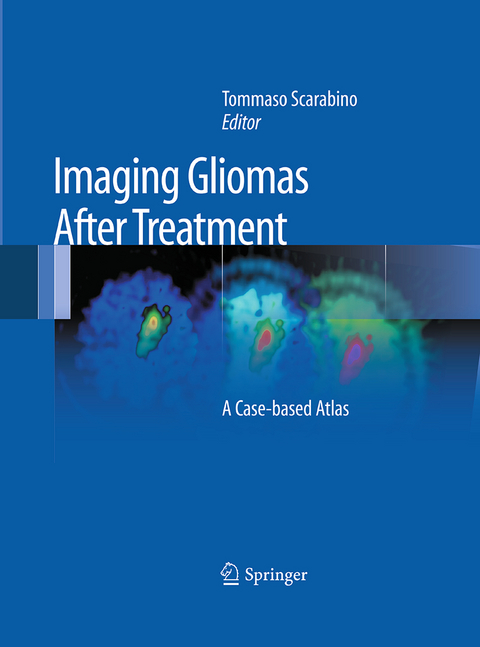
Imaging Gliomas After Treatment
A Case-based Atlas
Seiten
2016
|
Softcover reprint of the original 1st ed. 2012
Springer Verlag
978-88-470-5826-2 (ISBN)
Springer Verlag
978-88-470-5826-2 (ISBN)
This atlas is a detailed guide to the imaging appearances of gliomas following treatment with neurosurgery, radiation therapy, and chemotherapy. Particular emphasis is placed on characteristic appearances on the newer functional MR imaging techniques, including MR spectroscopy, diffusion-weighted imaging, and perfusion imaging.
This atlas is a detailed guide to the imaging appearances of gliomas following treatment with neurosurgery, radiation therapy, and chemotherapy. Normal and pathological findings are displayed in detailed MR images that illustrate the potential modifications due to treatment. Particular emphasis is placed on characteristic appearances on the newer functional MR imaging techniques, including MR spectroscopy, diffusion-weighted imaging, and perfusion imaging. These techniques are revolutionizing neuroradiology by going beyond the demonstration of macroscopic alterations to the depiction of preceding metabolic changes at the cellular and subcellular level, thereby allowing earlier and more specific diagnosis. A key section comprising some 40 clinical cases and more than 500 illustrations offers an invaluable clinical and research tool not only for neuroradiologists but also for neurosurgeons, radiotherapists, and medical oncologists.
This atlas is a detailed guide to the imaging appearances of gliomas following treatment with neurosurgery, radiation therapy, and chemotherapy. Normal and pathological findings are displayed in detailed MR images that illustrate the potential modifications due to treatment. Particular emphasis is placed on characteristic appearances on the newer functional MR imaging techniques, including MR spectroscopy, diffusion-weighted imaging, and perfusion imaging. These techniques are revolutionizing neuroradiology by going beyond the demonstration of macroscopic alterations to the depiction of preceding metabolic changes at the cellular and subcellular level, thereby allowing earlier and more specific diagnosis. A key section comprising some 40 clinical cases and more than 500 illustrations offers an invaluable clinical and research tool not only for neuroradiologists but also for neurosurgeons, radiotherapists, and medical oncologists.
Introduction.- Classification.- Gliomas.- Etiology – Heredity – Risk factors – Pathogenesis – Prognostic factors.- Complications – Signs and symptoms – Diagnosis and follow-up – Supportive therapy.- Treatment of gliomas.- Surgery – Radiotherapy – Chemotherapy.- Post-treatment neuroradiological imaging.-Morphological magnetic resonance.- Functional magnetic resonance: Spectroscopy – Diffusion – Perfusion – Cortical activation.- Clinical cases.- Bibliography.
| Erscheinungsdatum | 16.01.2018 |
|---|---|
| Zusatzinfo | XVIII, 201 p. |
| Verlagsort | Milan |
| Sprache | englisch |
| Maße | 193 x 260 mm |
| Themenwelt | Medizinische Fachgebiete ► Chirurgie ► Neurochirurgie |
| Medizin / Pharmazie ► Medizinische Fachgebiete ► Neurologie | |
| Medizin / Pharmazie ► Medizinische Fachgebiete ► Onkologie | |
| Medizinische Fachgebiete ► Radiologie / Bildgebende Verfahren ► Radiologie | |
| Schlagworte | fMRI • MR diffusion • MR perfusion • MR spectroscopy • Post-treatment MR imaging |
| ISBN-10 | 88-470-5826-0 / 8847058260 |
| ISBN-13 | 978-88-470-5826-2 / 9788847058262 |
| Zustand | Neuware |
| Informationen gemäß Produktsicherheitsverordnung (GPSR) | |
| Haben Sie eine Frage zum Produkt? |
Mehr entdecken
aus dem Bereich
aus dem Bereich
Buch | Hardcover (2024)
De Gruyter (Verlag)
CHF 153,90
Buch | Hardcover (2023)
Springer (Verlag)
CHF 307,95


