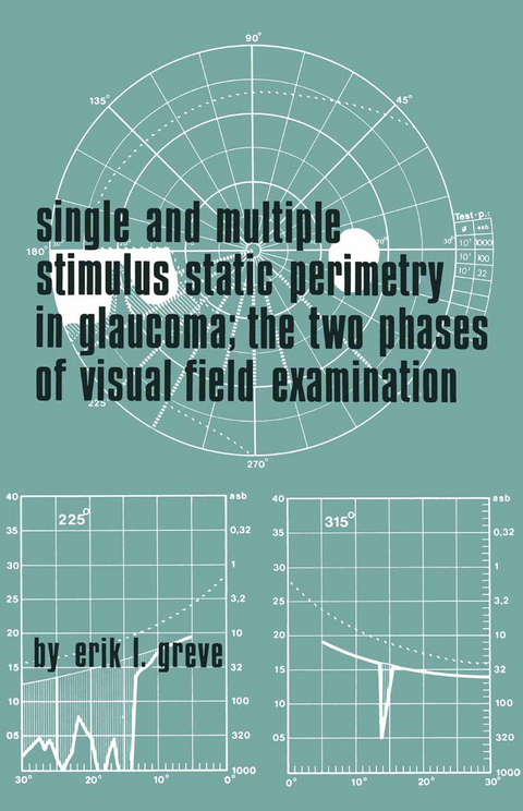
Single and Multiple Stimulus Static Perimetry in Glaucoma; The Two Phases of Perimetry
Kluwer Academic Publishers (Verlag)
9789061930020 (ISBN)
does not vary more than 1 % over the whole surface. Stimulus: The luminance of the stimuli can be regulated by means of neutral density filters. These filters are neither entirely neutral nor completely uniform, but these variations are not significant for perimetry. The maximum luminance of the stimuli is standardized at 1000 asb (316 2 cd.m-). The standard GP for kinetic perimetry has 3 neutral density filters which reduce the luminance by 0.5, 1.0 and 1.5 log units respectively. The modification of the GP for static perimetry is supplied with an additional series of four neutral density filters, which allow luminance steps of 0.1, 0.2, 0.3 and 0.4 log units. The resulting luminance values, expressed in apostilb, are given in Table I. TABLE I Luminance values of the Goldmann perimeter 4 3 2 steps lux 1430 450 143 45 3,1 asb 1000 315 100 31,5 3,1 2 cd/m 315 100 31,5 10 3,1 ßL ±30 10 3 3,1 L log L asb 3 2,5 2 2 1,5 0,5 2 Background: 31,5 asb. = 10 cd/m • Coefficient of reflection 0,7. 1 2 (1 asb = -cd/m).
I. Introduction.- II. Some Basic Facts from Visual Physiology.- Visual Field Examination is a threshold measurement.- Methods of measurement.- Factors which determine the difference threshold.- Physical characteristics of the stimulation.- Receptor and neural mechanisms.- III. The Fundamental Principles of Static and Kinetic Perimetry.- Static Perimetry.- Kinetic Perimetry.- The fundamental difference between static and kinetic perimetry.- Fundamental limits of the accuracy of kinetic perimetry.- Conclusion.- IV. Choice of the Conditions of Examination.- Choice of adaptation level.- Choice of stimulus size; spatial interaction and visual field examination.- Choice of presentation time; temporal interaction and visual field examination.- Local adaptation and visual field examination.- Some remarks about critical fusion frequency (C.F.F.) and colour in visual field examination.- V. The Place of Static and Kinetic Perimetry in the two Phases of Visual Field Examination.- Detection phase.- Assessment phase.- VI. Three Instruments for Single Stimulus Visual Field Examination.- The Goldmann perimeter.- The Tübinger perimeter.- The Double Projection Campimeter.- VII. Normal Values and Normal Variation of Thresholdmeasurements.- Normal values and inter-individual variation.- Intra-individual variation.- Influence of age.- Blind spot.- Angioscotomata.- Cartography and visual field.- VIII. Multiple Static Stimuli in the Detection Phase.- Specific problems of multiple stimuli.- Instruments for multiple stimulus visual field examination.- The Visual Field Analyser in the detection phase.- Optimum visual field examination using multiple stimuli.- Two other detection methods.- IX. Preretinal Factors.- Pupil.- Lens.- Ametropia.- Accommodation.- X. Description of Visual FieldDefects.- Topography.- Intensity.- XI. The Examination Procedure in Practice; Experiences with the Multiple Stimulus Method for the Detection of Glaucomatous Defects.- Examination procedure.- Glaucomatous defects.- Demonstration of 18 visual fields with glaucomatous defects.- Results of the comparative study.- Conclusion.- XII. Summary and conclusion.- References.
| Erscheint lt. Verlag | 30.6.1973 |
|---|---|
| Zusatzinfo | 368 p. |
| Verlagsort | Dordrecht |
| Sprache | englisch |
| Maße | 155 x 235 mm |
| Themenwelt | Medizin / Pharmazie ► Medizinische Fachgebiete ► Augenheilkunde |
| ISBN-13 | 9789061930020 / 9789061930020 |
| Zustand | Neuware |
| Informationen gemäß Produktsicherheitsverordnung (GPSR) | |
| Haben Sie eine Frage zum Produkt? |
aus dem Bereich


