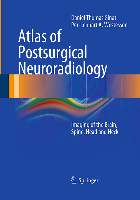
Atlas of Postsurgical Neuroradiology
Springer Berlin (Verlag)
978-3-662-50673-8 (ISBN)
As a result of the increasing number of surgical procedures on the brain, head, neck, and spine, postoperative changes are being encountered more frequently on neuroradiological examinations. However, these findings are often unfamiliar to neuroradiologists and neurosurgeons and can be difficult to interpret. This book, which contains numerous images and to-the-point case descriptions, is a comprehensive yet concise reference guide to postsurgical neuroradiology. It will enable the reader to identify the type of surgery performed and the hardware implanted and to differentiate expected sequelae from complications. Topics reviewed include trauma, tumors, vascular disorders, and infections of the head, neck, and spine; cerebrospinal fluid abnormalities; and degenerative diseases of the spine. This book will serve as a unique and convenient resource for both neuroradiologists and neurosurgeons.
Dr. Daniel T. Ginat works at the Division of Diagnostic and Interventional Neuroradiology in the Department of Imaging Sciences, University of Rochester School of Medicine and Dentistry, where he has three times won the RAIN (Resident Achievement in Neuroradiology) award. Dr. Ginat is the recipient of a Harry W. Fischer Research Fund Grant and has also received a Roentgen Resident/Fellow Research Award from the Radiological Society of North America. Professor Per-Lennart Westesson is Director of the Division of Diagnostic and Interventional Neuroradiology at the University of Rochester School of Medicine and Dentistry. Professor Westesson initially studied dentistry and subsequently obtained board certification in diagnostic radiology. He is a highly respected expert in the field. His publications include more than 180 journal articles and well-received books on maxillofacial imaging and diffusion-weighted imaging of the brain. Professor Westesson is the recipient of numerous awards, including the Magna Cum Laude Award from the American Society of Neuroradiology.
Introduction.- Brain: Burr holes/craniotomy/craniectomy/tumor resection. Devices.- Skull base: Anterior craniofacial resection. Pituitary tumor resection. Craniopharyngioma resection. Suprasellar cyst fenestration. Encephalocele repair. Temporal bone. Complications.- Craniofacial: Craniosynostosis repair. LeFort osteotomy. Sagittal split. Fracture repair. Septoplasty. Cosmesis. Complications.- Head and Neck: Neck. Mandible. Pharynx. Orbit. Flap reconstruction. Thyroidectomy. Parathyroidectomy. Paranasal sinuses. Devices.- Spine: Variety of procedures and hardware. Spine stimulation. Kyphoplasty/vertebroplasty/sacroplasty. Failed back surgery syndrome.- Vascular: Variety of clips, coils, stents. Aneurysm treatment. AVM/AVF/covernoma treatment. Embolectomy. Angioplasty. Synangiosis. Venous sinus skeletonization. CEA. Carotid-axillary bypass. Complications.- CSF shunts: Ventriculoperitoneal shunts. Lumboperitoneal shunts.
From the reviews:
"This monograph concerns the postoperative findings using CT, MRI, PET, and plain radiographic means to evaluate head, neck, skull, and brain surgeries. I highly recommend this book with its great depth and scope for neurosurgeons seeking their validation postoperatively for minor to major techniques. This is meant to help clinical neurosurgeons with routine patient followup and evaluation." (Joseph J. Grenier, Amazon.com, February, 2014)
"It fills a gap in the literature and can serve as a handy reference for practicing radiologists. The target audience consists of residents rotating in neuroradiology, neuroradiology fellows, and practicing neuroradiologists. ... This is a body of work that covers a broad range of common and uncommon surgical procedures, surgical hardware, and postsurgical complications. It is a useful reference for radiologists at varying levels of training or years of practice. There is no doubt that it will find its place near workstations throughout the world." (Scott E. Forseen, Doody's Book Reviews, May, 2013)
| Erscheinungsdatum | 19.08.2017 |
|---|---|
| Zusatzinfo | XX, 653 p. |
| Verlagsort | Berlin |
| Sprache | englisch |
| Maße | 178 x 254 mm |
| Gewicht | 1267 g |
| Themenwelt | Medizinische Fachgebiete ► Chirurgie ► Neurochirurgie |
| Medizin / Pharmazie ► Medizinische Fachgebiete ► HNO-Heilkunde | |
| Medizinische Fachgebiete ► Radiologie / Bildgebende Verfahren ► Radiologie | |
| Medizin / Pharmazie ► Studium | |
| Schlagworte | complications • devices • Head and Neck Surgery • Imaging • Neuroradiology • neurosurgery |
| ISBN-10 | 3-662-50673-4 / 3662506734 |
| ISBN-13 | 978-3-662-50673-8 / 9783662506738 |
| Zustand | Neuware |
| Informationen gemäß Produktsicherheitsverordnung (GPSR) | |
| Haben Sie eine Frage zum Produkt? |
aus dem Bereich


