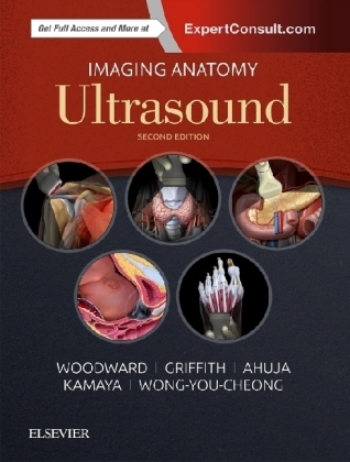
Imaging Anatomy: Ultrasound
Elsevier - Health Sciences Division (Verlag)
978-0-323-54800-7 (ISBN)
- Titel ist leider vergriffen;
keine Neuauflage - Artikel merken
Key Features:
- Provides expert reference at the point of care in every anatomical area where ultrasound is used
- Presents richly labeled images with associated commentary as well as thumbnail scout images to show transducer placement
- Features a robust collection of CT/MR correlations, highlighting the importance of multimodality imaging in modern clinical practice
Paula J. Woodward, Professor of Radiology, David G. Bragg, MD and Marcia R. Bragg Presidential Endowed Chair in Oncologic Imaging, Adjunct Professor of Obstetrics and Gynecology, University of Utah School of Medicine, Salt Lake City, Utah
James F. Griffith, Research Assistant Professor, Northwestern University. Dr. Griffith’s area of expertise is applying measurement science and psychometrics to clinical problems. He has extensive experience both researching and teaching in this area. He’s additionally provided consultation on the topic to a variety of non-profits, pharmaceutical, and medical device companies. He maintains a clinical practice in Chicago focused on cognitive behavioral therapy for adults with anxiety and depression. He has published over 70 papers on various aspects of psychometrics and psychological assessment.
Gregory Antonio, Honorary Professor, Department of Imaging and Interventional Radiology, The Chinese University of Hong Kong, Consultant Radiologist, Scanning Department, St. Teresa’s Hospital, Hong Kong (SAR), China
Anil T. Ahuja, Professor of Diagnostic Radiology & Organ Imaging, Faculty of Medicine, The Chinese University of Hong Kong, Prince of Wales Hospital, Hong Kong (SAR), China
K. Wong, Consultant & Clinical Associate Professor (Honorary), Department of Imaging and Interventional Radiology, Prince of Wales Hospital, Faculty of Medicine, The Chinese University of Hong Kong, Hong Kong (SAR), China
Aya Kamaya, Associate Professor of Radiology, Director, Stanford Body Imaging Fellowship, Stanford University School of Medicine, Stanford, California
Jade Wong-You-Cheong, Professor, Department of Diagnostic Radiology and Nuclear Medicine, University of Maryland School of Medicine, Director of Ultrasound, University of Maryland Medical Center, Baltimore, Maryland
BRAIN AND SPINE
Scalp and Calvarial Vault
Cranial Meninges
Cerebral Hemispheres Overview
Brainstem and Cerebellum
Midbrain
Ventricles and Choroid Plexus
Subarachnoid Space/Cisterns
Orbit
Trans-Cranial Color Doppler Overview
Anterior Cerebral Artery
Middle Cerebral Artery
Posterior Cerebral Artery
Distal Internal Carotid Artery
Vertebrobasilar System
Intracranial Veins
Vertebral Bodies, Spinal Cord & Cauda Equina
NECK View less >
Neck Overview
Sublingual/Submental Region
Submandibular Region
Parotid Region
Upper Cervical Level
Midcervical Level
Lower Cervical Level and Supraclavicular Fossa
Posterior Triangle
Thyroid Gland
Parathyroid Gland
Larynx and Hypopharynx
Cervical Trachea and Esophagus
Brachial Plexus
Vagus Nerve
Cervical Carotid Arteries
Vertebral Arteries
Neck Veins
Cervical Lymph Nodes
THORAX View less >
Thoracic Outlet
Pleura
Diaphragm
Ribs and Intercostal Space
Breast
ABDOMEN View less >
Liver
Biliary System
Spleen
Pancreas
Kidneys
Adrenals
Bowel
Abdominal Lymph Nodes
Aorta and Inferior Vena Cava
Peritoneal Spaces and Structures
Abdominal Wall
PELIVS View less >
Ureters and Bladder
Prostate
Testes
Penis and Male Urethra
Uterus
Cervix
Vagina
Ovaries
Iliac Arteries and Veins
Trans-Perineal Anatomy
UPPER LIMB View less >
Sternoclavicular and Acromioclavicular Joints
Shoulder
Axilla
Arm
Arm Vessels
Elbow
Forearm
Forearm Vessels
Wrist
Hand
Hand Vessels
Thumb
Fingers
Radial Nerve
Median Nerve
Ulnar Nerve
LOWER LIMB
Hip
Thigh Muscles
Femoral Vessels and Nerves
Knee
Leg Muscles
Leg Vessels
Leg Nerves
Ankle
Tarsus
Foot Vessels
Metatarsals and Toes
OBSTETRICS
Embryology and Anatomy of the First Trimester
Embryology and Anatomy of the Brain
Embryology and Anatomy of the Spine
Embryology and Anatomy of the Face and Neck
Embryology and Anatomy of the Chest
Embryology and Anatomy of the Cardiovascular System
Embryology and Anatomy of the Abdominal Wall and GI Tract
Embryology and Anatomy of the Genitourinary Tract
| Erscheinungsdatum | 28.11.2017 |
|---|---|
| Zusatzinfo | Approx. 2200 illustrations (2200 in full color) |
| Verlagsort | Philadelphia |
| Sprache | englisch |
| Maße | 222 x 281 mm |
| Gewicht | 3080 g |
| Einbandart | gebunden |
| Themenwelt | Medizin / Pharmazie ► Medizinische Fachgebiete ► Onkologie |
| Medizinische Fachgebiete ► Radiologie / Bildgebende Verfahren ► Sonographie / Echokardiographie | |
| Studium ► 1. Studienabschnitt (Vorklinik) ► Anatomie / Neuroanatomie | |
| ISBN-10 | 0-323-54800-8 / 0323548008 |
| ISBN-13 | 978-0-323-54800-7 / 9780323548007 |
| Zustand | Neuware |
| Informationen gemäß Produktsicherheitsverordnung (GPSR) | |
| Haben Sie eine Frage zum Produkt? |
aus dem Bereich


