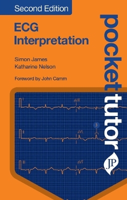
Pocket Tutor ECG Interpretation
JP Medical Ltd (Verlag)
978-1-909836-71-6 (ISBN)
Topics reflect information needs stemming from today’s integrated undergraduate and foundation courses:
Common presentations
Investigation options (e.g. ECG, imaging)
Clinical and patient-orientated skills (e.g. examinations, history-taking)
The highly-structured, bite-size content helps novices combat the ‘fear factor’ associated with day-to-day clinical training, and provides a detailed resource that students and junior doctors can carry in their pocket.
Key points
New edition of the best-selling title that breaks down a complex and daunting subject using clearly-labelled, full-page ECG traces and concise but informative text
Revised text and brand-new ECG traces bring the new edition fully up-to-date
New chapters cover electrolyte and homeostatic disorders, and normal variants
Logical, sequential content: relevant basic science, then a guide to understanding a normal ECG and the building blocks of an abnormal ECG, before describing clinical disorders
Simon James MBBS MRCP Consultant Electrophysiologist, James Cook University Hospital, Middlesbrough, UK Katharine Nelson MBBS MRCP Consultant in Cardiology, Queen Elizabeth Hospital, Gateshead Health NHS Foundation Trust, Gateshead, UK
Chapter 1 First principles
1.1 Anatomy
1.2 Physiology
1.3 Electrical activity and the ECG
Chapter 2 Understanding the normal ECG
2.1 Introduction
2.2 The limb leads
2.3 The chest leads
2.4 The lead orientation
2.5 ECG nomenclature
Chapter 3 Interpreting the ECG: a six-step approach
3.1 Step 1: is there electrical activity?
3.2 Step 2: what is the QRS (ventricular) rate?
3.3 Step 3: is the rhythm regular?
3.4 Step 4: is the QRS narrow (normal) or broad?
3.5 Step 5: is there atrial electrical activity?
3.6 Step 6: how is the atrial activity related to the ventricular activity?
3.7 Glossary of distinct ECG signs
Chapter 4 Bradyarrhythmias I: sinoatrial node dysfunction
4.1 Sinus bradycardia
4.2 Sinus pause with junctional escape beat
Chapter 5 Bradyarrhythmias II: conduction abnormalities
5.1 First-degree atrioventricular block
5.2 Second-degree atrioventricular block: Mobitz type 1 or Wenckebach
5.3 Second-degree atrioventricular block: Mobitz type 2
5.4 Second-degree heart block: 2:1 atrioventricular block
5.5 Third-degree (complete) atrioventricular block: narrow QRS
5.6 Third-degree (complete) atrioventricular block: broad QRS
5.7 Right bundle branch block
5.8 Left bundle branch block
Chapter 6 Ectopic beats
6.1 Atrial ectopic beats
6.2 Ventricular ectopic beats
6.3 Junctional ectopic beats
Chapter 7 Atrial arrhythmias
7.1 Atrial tachycardia
7.2 Multifocal atrial tachycardia
7.3 Atrial flutter
7.4 Atrial fibrillation
7.5 Atrial fibrillation with left bundle branch block
Chapter 8 Narrow-complex tachyarrhythmias (supraventricular tachycardias)
8.1 Atrioventricular nodal re-entrant tachycardia
8.2 Atrioventricular reciprocating tachycardia
8.3 Wolff–Parkinson–White syndrome: right-sided pathway
8.4 Wolff–Parkinson–White syndrome: left lateral pathway
8.5 Wolff–Parkinson–White syndrome: posterior pathway
8.6 Wolff–Parkinson–White syndrome plus atrial fibrillation
Chapter 9 Broad-complex tachyarrhythmias
9.1 Monomorphic ventricular tachycardia
9.2 Polymorphic ventricular tachycardia
9.3 Torsade de points
9.4 Ventricular fibrillation
9.5 Supraventricular tachycardia with bundle branch block
Chapter 10 Ischaemia and infarction
10.1 ST segment depression (cardiac ischaemia)
10.2 Acute myocardial ischaemia: T wave inversion and the LAD syndrome
10.3 ST segment elevation myocardial infarction: anterior0
10.4 ST segment elevation myocardial infarction: acute inferior
10.5 ST segment elevation myocardial infarction: posterior
10.6 Completed myocardial infarction
Chapter 11 Inherited arrhythmia problems
11.1 Hypertrophic cardiomyopathy
11.2 Arrhythmogenic right ventricular dysplasia
11.3 Long QT syndrome
11.4 Brugada syndrome
Chapter 12 Cardiac chamber dilation, strain and hypertrophy
12.1 Left ventricular hypertrophy
12.2 Right ventricular hypertrophy
12.3 Pulmonary embolus
12.4 Left atrial dilation
12.5 Right atrial dilation
Chapter 13 Electrolyte and homeostasis disorders
13.1 Hyperkalaemia
13.2 Hypokalaemia
13.3 Hypercalcaemia
13.4 Hypocalcaemia
13.5 Hypothermia
Chapter 14 Miscellaneous conditions and normal variants
14.1 Pericarditis
14.2 Pacemaker
14.3 Biventricular pacemaker
14.4 Early repolarisation pattern
14.5 Non-pathological Q waves
| Erscheinungsdatum | 11.09.2019 |
|---|---|
| Verlagsort | London |
| Sprache | englisch |
| Maße | 113 x 177 mm |
| Themenwelt | Medizinische Fachgebiete ► Innere Medizin ► Kardiologie / Angiologie |
| Medizin / Pharmazie ► Medizinische Fachgebiete ► Radiologie / Bildgebende Verfahren | |
| ISBN-10 | 1-909836-71-0 / 1909836710 |
| ISBN-13 | 978-1-909836-71-6 / 9781909836716 |
| Zustand | Neuware |
| Haben Sie eine Frage zum Produkt? |
aus dem Bereich


