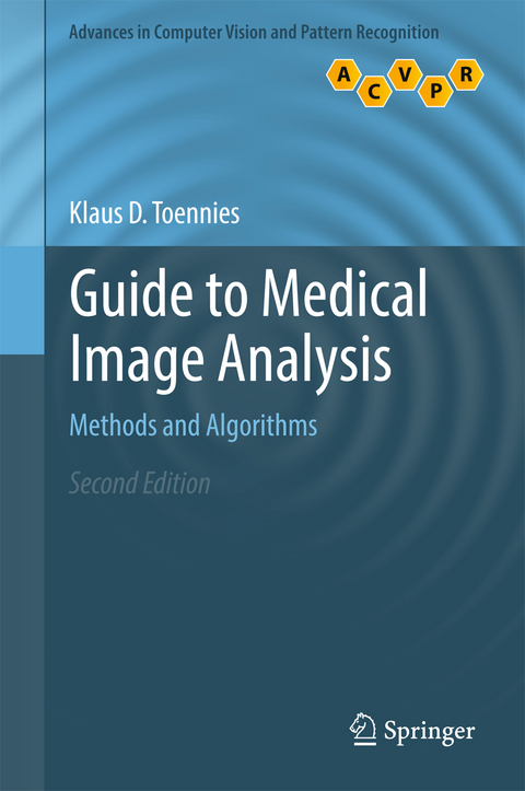
Guide to Medical Image Analysis
Methods and Algorithms
Seiten
2017
|
2nd ed. 2017
Springer London Ltd (Verlag)
978-1-4471-7318-2 (ISBN)
Springer London Ltd (Verlag)
978-1-4471-7318-2 (ISBN)
This comprehensive guide provides a uniquely practical, application-focused introduction to medical image analysis. describes a range of common imaging techniques, reconstruction techniques and image artifacts, and discusses the archival and transfer of images;
This comprehensive guide provides a uniquely practical, application-focused introduction to medical image analysis. This fully updated new edition has been enhanced with material on the latest developments in the field, whilst retaining the original focus on segmentation, classification and registration. Topics and features: presents learning objectives, exercises and concluding remarks in each chapter; describes a range of common imaging techniques, reconstruction techniques and image artifacts, and discusses the archival and transfer of images; reviews an expanded selection of techniques for image enhancement, feature detection, feature generation, segmentation, registration, and validation; examines analysis methods in view of image-based guidance in the operating room (NEW); discusses the use of deep convolutional networks for segmentation and labeling tasks (NEW); includes appendices on Markov random field optimization, variational calculus and principal component analysis.
This comprehensive guide provides a uniquely practical, application-focused introduction to medical image analysis. This fully updated new edition has been enhanced with material on the latest developments in the field, whilst retaining the original focus on segmentation, classification and registration. Topics and features: presents learning objectives, exercises and concluding remarks in each chapter; describes a range of common imaging techniques, reconstruction techniques and image artifacts, and discusses the archival and transfer of images; reviews an expanded selection of techniques for image enhancement, feature detection, feature generation, segmentation, registration, and validation; examines analysis methods in view of image-based guidance in the operating room (NEW); discusses the use of deep convolutional networks for segmentation and labeling tasks (NEW); includes appendices on Markov random field optimization, variational calculus and principal component analysis.
Dr. Klaus D. Toennies is a Professor of Image Processing and Pattern Recognition at the Department of Simulation and Graphics of the Otto-von-Guericke University of Magdeburg, Germany.
The Analysis of Medical Images.- Digital Image Acquisition.- Image Storage and Transfer.- Image Enhancement.- Feature Detection.- Segmentation: Principles and Basic Techniques.- Segmentation in Feature Space.- Segmentation as a Graph Problem.- Active Contours and Active Surfaces.- Registration and Normalization.- Shape, Appearance and Spatial Relationships.- Classification and Clustering.- Validation.- Appendix.
| Erscheinungsdatum | 16.05.2017 |
|---|---|
| Reihe/Serie | Advances in Pattern Recognition |
| Zusatzinfo | 197 Illustrations, color; 187 Illustrations, black and white; XXIV, 589 p. 384 illus., 197 illus. in color. |
| Verlagsort | England |
| Sprache | englisch |
| Maße | 155 x 235 mm |
| Themenwelt | Informatik ► Grafik / Design ► Digitale Bildverarbeitung |
| Informatik ► Theorie / Studium ► Künstliche Intelligenz / Robotik | |
| Medizinische Fachgebiete ► Radiologie / Bildgebende Verfahren ► Radiologie | |
| ISBN-10 | 1-4471-7318-X / 144717318X |
| ISBN-13 | 978-1-4471-7318-2 / 9781447173182 |
| Zustand | Neuware |
| Haben Sie eine Frage zum Produkt? |
Mehr entdecken
aus dem Bereich
aus dem Bereich
alles zum Drucken, Scannen, Modellieren
Buch | Softcover (2024)
Markt + Technik Verlag
CHF 34,90
Methoden, Konzepte und Algorithmen in der Optotechnik, optischen …
Buch | Hardcover (2024)
Hanser (Verlag)
CHF 55,95


