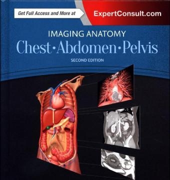
Imaging Anatomy: Chest, Abdomen, Pelvis
Elsevier - Health Sciences Division (Verlag)
978-0-323-47781-9 (ISBN)
- Titel erscheint in neuer Auflage
- Artikel merken
Includes all relevant imaging modalities, 3D reconstructions, and highly accurate and detailed medical drawings that illustrate the fine points of the imaging anatomy
Depicts common anatomic variants and covers common pathological processes as a part of its comprehensive coverage
Provides a detailed overview of airway and interstitial network anatomy-the basis for understanding and diagnosing interstitial lung disease
Features representative pathologic examples to highlight the effect of disease on human anatomy
Includes plain radiography, the latest generation of multi-planar advanced cross-sectional MR and CT, ultrasound for pelvis/renal/liver/gallbladder, barium for GI tract, and much more
Offers state of the art, detailed pelvic floor imaging and perianal/perirectal fistula imaging using high-resolution CT and MR, including 3T MR
Expert Consult eBook version included with purchase. This enhanced eBook experience allows you to search all of the text, figures, images, and references from the book on a variety of devices.
Michael P. Federle, MD, FACR, Professor and Associate Chair for Education, Department of Radiology, Stanford University School of Medicine Stanford, California Melissa L. Rosado-de-Christenson, MD, FACR, FAAWR, is Attending Radiologist at the Division of Cardiothoracic Imaging in the Department of Medical Imaging for Banner - University Medical Group Tucson,. She is also Professor of Medical Imaging at University of Arizona College of Medicine - Tucson, in Tucson, Arizona Dr. Siva Raman is a board-certified radiologist who gained his subspecialty expertise in thoracoabdominal imaging during a fellowship at Stanford University, and intern residency at UC Davis Medical Center. He attended Johns Hopkins University Medical School. Dr. Raman is a radiologist at Bay Imaging Consultants in Walnut Creek, California, with a subspecialty focus on thoracoabdominal imaging Paula J. Woodward MD is a Professor in the Department of Radiology and Adjunct Professor of Obstetrics and Gynecology at the University of Utah. Dr. Woodward is a practicing diagnostic radiologist who specializes in CT, MR, US, and x-ray imaging modalities as well as obstetrical ultrasound and GYN imaging, and she holds the David G. Bragg, MD and Marcia R. Bragg Presidential Endowed Chair in Oncologic Imaging. Dr Woodward completed her residency training at Wilford Hall USAF Medical Center.
CHEST
Chest Overview
Lung Development
Airway Structure
Vascular Structure
Interstitial Network
Lungs
Hila
Airways
Pulmonary Vessels
Pleura
Mediastinum
Systemic Vessels
Heart
Coronary Arteries and Cardiac Veins
Pericardium
Chest Wall
ABDOMEN
Embryology of Abdomen
Abdominal Wall
Diaphragm
Peritoneal Cavity
Vessels, Lymphatic System and Nerves, Abdominal
Esophagus
Gastroduodenal
Small Intestine
Colon
Spleen
Liver
Biliary System
Pancreas
Retroperitoneum
Adrenal
Kidney
Ureter and Bladder
PELVIS
Vessels, Lymphatic System and Nerves, Pelvic
Male
Male Pelvic Wall and Floor
Testes and Scrotum
Prostate and Seminal Vesicles
Penis and Urethra
Female
Female Pelvic Floor
Uterus
Ovaries
| Erscheinungsdatum | 23.12.2016 |
|---|---|
| Reihe/Serie | Imaging Anatomy |
| Verlagsort | Philadelphia |
| Sprache | englisch |
| Maße | 216 x 276 mm |
| Gewicht | 3580 g |
| Themenwelt | Medizinische Fachgebiete ► Radiologie / Bildgebende Verfahren ► Radiologie |
| Studium ► 1. Studienabschnitt (Vorklinik) ► Anatomie / Neuroanatomie | |
| ISBN-10 | 0-323-47781-X / 032347781X |
| ISBN-13 | 978-0-323-47781-9 / 9780323477819 |
| Zustand | Neuware |
| Haben Sie eine Frage zum Produkt? |
aus dem Bereich


