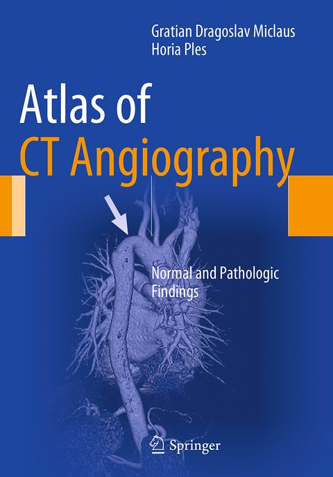
Atlas of CT Angiography
Normal and Pathologic Findings
Seiten
2016
|
Softcover reprint of the original 1st ed. 2014
Springer International Publishing (Verlag)
978-3-319-35688-4 (ISBN)
Springer International Publishing (Verlag)
978-3-319-35688-4 (ISBN)
This superbly illustrated atlas presents CT angiography with 3D reconstruction in a broad range of clinical applications. Includes imaging of cerebral, carotid, thoracic, coronary, abdominal and peripheral vessels, comparing normal and pathologic appearances.
This atlas presents normal and pathologic findings observed on CT angiography with 3D reconstruction in a diverse range of clinical applications, including the imaging of cerebral, carotid, thoracic, coronary, abdominal and peripheral vessels. The superb illustrations display the excellent anatomic detail obtained with CT angiography and depict the precise location of affected structures and lesion severity. Careful comparisons between normal imaging features and pathologic appearances will assist the reader in image interpretation and treatment planning and the described cases include some very rare pathologies. In addition, the technical principles of the modality are clearly explained and guidance provided on imaging protocols. This atlas will be of value both to those in training and to more experienced practitioners within not only radiology but also cardiovascular surgery, neurosurgery, cardiology and neurology.
This atlas presents normal and pathologic findings observed on CT angiography with 3D reconstruction in a diverse range of clinical applications, including the imaging of cerebral, carotid, thoracic, coronary, abdominal and peripheral vessels. The superb illustrations display the excellent anatomic detail obtained with CT angiography and depict the precise location of affected structures and lesion severity. Careful comparisons between normal imaging features and pathologic appearances will assist the reader in image interpretation and treatment planning and the described cases include some very rare pathologies. In addition, the technical principles of the modality are clearly explained and guidance provided on imaging protocols. This atlas will be of value both to those in training and to more experienced practitioners within not only radiology but also cardiovascular surgery, neurosurgery, cardiology and neurology.
Cerebral angiography.- Carotid angiography.- Thoracic angiography.- Coronary angiography.- Abdominal angiography.- Peripheral angiography.
| Erscheinungsdatum | 03.08.2016 |
|---|---|
| Zusatzinfo | XI, 198 p. 489 illus., 355 illus. in color. |
| Verlagsort | Cham |
| Sprache | englisch |
| Maße | 178 x 254 mm |
| Themenwelt | Medizinische Fachgebiete ► Innere Medizin ► Kardiologie / Angiologie |
| Medizin / Pharmazie ► Medizinische Fachgebiete ► Neurologie | |
| Medizinische Fachgebiete ► Radiologie / Bildgebende Verfahren ► Radiologie | |
| Schlagworte | aneurisms • Cardiology • Cardiovascular medicine • Carotid Imaging • Cerebral Imaging • Congenital malformations • CT angiography • Imaging / Radiology • Medical Imaging • Medicine • Neurology • Neurology and clinical neurophysiology • Radiology • Stenosis |
| ISBN-10 | 3-319-35688-7 / 3319356887 |
| ISBN-13 | 978-3-319-35688-4 / 9783319356884 |
| Zustand | Neuware |
| Informationen gemäß Produktsicherheitsverordnung (GPSR) | |
| Haben Sie eine Frage zum Produkt? |
Mehr entdecken
aus dem Bereich
aus dem Bereich
in Fällen, Fragen und Antworten
Buch | Softcover (2024)
Urban & Fischer in Elsevier (Verlag)
CHF 124,60
Diagnostik und interventionelle Therapie | 2 Bände
Buch (2024)
Deutscher Ärzteverlag
CHF 489,95
Buch | Softcover (2023)
Urban & Fischer in Elsevier (Verlag)
CHF 61,60


