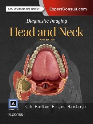
Diagnostic Imaging: Head and Neck
Seiten
2016
|
3rd edition
Elsevier - Health Sciences Division (Verlag)
978-0-323-44301-2 (ISBN)
Elsevier - Health Sciences Division (Verlag)
978-0-323-44301-2 (ISBN)
- Titel erscheint in neuer Auflage
- Artikel merken
Zu diesem Artikel existiert eine Nachauflage
Nearly 400 diagnoses that are delineated, referenced, and lavishly illustrated highlight the third edition of this bestselling reference. Dr. H. Ric Harnsberger and his expert author team of Drs. Pat Hudgins, Bernadette L. Koch, and Bronwyn Hamilton provide carefully updated information in a concise, bulleted format, keeping you current with recent advances in head and neck radiology. Succinct text, outstanding illustrations, and up-to-date content make this title a must-have reference for both radiologists and otolaryngologists who need a single, go-to guide in this fast-changing area.
Concise, bulleted text provides efficient information on nearly 400 diagnoses that are clearly illustrated with over 2800 superb images
Designed for quick and easy clinical reference at the point of care, with logically organized sections, comprehensive lists of differential diagnosis, consistent presentation of information, and relevant, newly revised images throughout
Expert ConsultT eBook version included with purchase, which allows you to search all of the text, figures, and references from the book on a variety of devices
Meticulously updated throughout, with new diagnoses, sequential imaging examples, and high-resolution 3T MR and 0.6-mm CT scans that provide the most current information in the field
New chapters on temporal bone and skull base tumors, as well as updated primary tumor staging (T) and nearby lymph node staging (N), keep you up-to-date
| Erscheinungsdatum | 29.11.2016 |
|---|---|
| Reihe/Serie | Diagnostic Imaging |
| Zusatzinfo | Approx. 3000 illustrations (3000 in full color); Illustrations |
| Verlagsort | Philadelphia |
| Sprache | englisch |
| Maße | 222 x 281 mm |
| Gewicht | 3960 g |
| Themenwelt | Medizinische Fachgebiete ► Radiologie / Bildgebende Verfahren ► Radiologie |
| ISBN-10 | 0-323-44301-X / 032344301X |
| ISBN-13 | 978-0-323-44301-2 / 9780323443012 |
| Zustand | Neuware |
| Informationen gemäß Produktsicherheitsverordnung (GPSR) | |
| Haben Sie eine Frage zum Produkt? |
Mehr entdecken
aus dem Bereich
aus dem Bereich
Buch (2023)
Thieme (Verlag)
CHF 265,95


