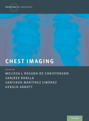
Chest Imaging
Oxford University Press Inc (Verlag)
978-0-19-985806-4 (ISBN)
Part of the Rotations in Radiology series, this book offers a guided approach to imaging diagnosis with examples of all imaging modalities complimented by the basics of interpretation and technique and the nuances necessary to arrive at the best diagnosis. Each chapter contains a targeted discussion of a pathology which reviews the definition, clinical features, anatomy and physiology, imaging techniques, differential diagnosis, clinical issues, key points, and further reading. This book is a must-read for residents and practitioners in radiology seeking refreshing on essential facts and imaging abnormalities in thoracic imaging.
Melissa L Rosado-de-Christenson is a Professor of Clinical Radiology and the Section Chief for Thoracic Radiology at the University of Missouri-Kansas City, St. Luke's Hospital in Kansas City, Missouri. Sanjeev Bhalla is an Associate Professor of Radiology at Washington University and the Chief of Cardiothoracic Imaging at the Mallinckrodt Institute of Radiology in St. Louis, Missouri. Gerald Abbott is an Associate Radiologist in the Division of Thoracic Imaging at Massachusetts General Hospital and Harvard Medical School in Boston, Massachusetts. Santiago Martinez-Jiminez is an Assistant Professor of Clinical Radiology at the University of Missouri-Kansas City, St. Luke's Hospital in Kansas City, Missouri.
Part I. Introduction to Chest Radiology
1. Introduction to Chest Radiology
2. Imaging Modalities
3. Overview of Normal Thoracic Imaging Anatomy
4. The radiology report and radiologic interpretation
5. Basic signs in chest radiology
Part II Bedside Radiography
6. Introduction to Portable Chest Radiography: Support Devices
7. Endotracheal and Enteric Tubes
8. Vascular Catheters
9. Pacers / AICD
10. Thoracostomy tubes, mediastinal drains
Part III Bedside Radiography - Common abnormalities
11 Bedside Chest Radiography-Common Findings
12. Pulmonary venous hypertension and edema
13. Airspace Disease - Atelectasis, pneumonia and aspiration
14. Acute respiratory distress syndrome
Part IV Volume Loss
15. Volume Loss
16. Atelectasis: Opaque hemithorax
17. Upper and middle lobe atelectasis
18. Lower lobe atelectasis
19. Subsegmental and rounded atelectasis
Part V Emergency Radiology
20. Introduction to Emergency Chest Radiology
21. Traumatic vascular injury
22. Chest trauma; Contusions, lacerations, fractures
23. Acute aortic syndromes and aortitis
24. Pulmonary thromboembolic disease
25. Sickle cell disease
26. Pulmonary hypertension
Part VI Pleural disease
27. Introduction to Pleural disease
28. Pneumothorax
29. Effusion
30. Empyema and complications
31. Pleural thickening and calcification
32. Pleural neoplasms
Part VII Pulmonary infections
33. Pulmonary infections
34. Community acquired pneumonia
35. Cavitary nodules, masses and consolidations
36. Endemic fungal pneumonias
37. Opportunistic fungal pneumonias
38. Tuberculosis
39. Non tuberculous mycobacterial infection
40. Mycoplasma and Viral pneumonias
Part VIII The immunocompromised patient
41. The immunocompromised patient
42. The immunocompromised patient - AIDS
43. The immunocompromised patient - Non-AIDS
Part IX Neoplasms of the lung and tracheobronchial tree
44. Introduction to neoplasms of lung and tracheobronchial tree
45. Lung cancer
46. Nodules and masses
47. Atelectasis and post-obstructive pneumonia
48. Lymphadenopathy and extrapulmonary involvement
49. Lung Cancer Staging
50. Bronchial carcinoid and bronchial gland carcinomas
51. Pulmonary metastases
52. Hamartoma and benign tumor-like lesions
Part X Airways Disease
53. Airways Disease
54. Tracheal narrowing and tracheomalacia
55. Bronchiectasis
56. Emphysema
57. Bronchiolitis
Part XI Connective Tissue Disorders and Autoimmune Conditions
58. Introduction to Connective Tissue Disorders and Autoimmune Conditions
59. Rheumatoid arthritis
60. Systemic Sclerosis
61. Pulmonary Vasculitis
62. Other autoimmune disorders
63. Eosinophilic lung diseases
64. Amyloidosis
Part XII Pneumoconiosis
65. Pneumoconiosis
66. Asbestosis
67. Silocosis and Coal Worker's Pneumoconiosis
Part XIII Iatrogenic conditions
68. Introduction to Iatrogenic Conditions
69. Radiation pneumonitis and fibrosis
70. Drug-induced lung disease
71. Post-operative complications
Part XIV Diffuse Infiltrative Lung Disease
72. Diffuse Infiltrative Lung Disease
73. Ground glass opacity
74. Cystic lung disease
75. Pulmonary micronodules
76. Sarcoidosis
Part XV Idiopathic interstitial pneumonias
77. Idiopathic interstitial pneumonias
78. Idiopathic pulmonary fibrosis
79. Non-Specific interstitial pneumonia
80. Cryptogenic organizing pneumonia
Part XVI Mediastinal Abnormalities
81. Introduction to Mediastinal Abnormalities
82. Primary neoplasms
83. Lymphadenopathy
84. Cysts
85. Glandular enlargement
86. Vascular lesions including lymphatic malformations and aneurysms
87. Miscellaneous, including herniations and EMH
Part XVII Developmental Abnormalities
88. Introduction to developmental abnormalities
89. Congenital bronchial atresia
90. Sequestration
91. Abnormal aorta and aortic branching
92. Abnormalities of pulmonary venous return
93. Abnornalities of systemic veins
94. Shunts including intracardiac and intrapulmonary
Part XVIII Diseases of the Chest Wall and Diaphragm
95. Diseases of the Chest Wall and Diaphragm
96. Chest wall abnormalities
97. Diaphragmatic abnormalities
| Erscheinungsdatum | 24.05.2016 |
|---|---|
| Reihe/Serie | Rotations in Radiology |
| Verlagsort | New York |
| Sprache | englisch |
| Maße | 282 x 223 mm |
| Gewicht | 2055 g |
| Themenwelt | Medizinische Fachgebiete ► Chirurgie ► Herz- / Thorax- / Gefäßchirurgie |
| Medizinische Fachgebiete ► Innere Medizin ► Pneumologie | |
| Medizinische Fachgebiete ► Radiologie / Bildgebende Verfahren ► Radiologie | |
| ISBN-10 | 0-19-985806-3 / 0199858063 |
| ISBN-13 | 978-0-19-985806-4 / 9780199858064 |
| Zustand | Neuware |
| Haben Sie eine Frage zum Produkt? |
aus dem Bereich


