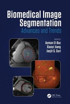
Biomedical Image Segmentation
Crc Press Inc (Verlag)
978-1-4822-5855-4 (ISBN)
Ayman El-Baz, Ph.D, is an associate professor in the Department of Bioengineering at the University of Louisville, Kentucky, USA. Dr. El-Baz has twelve years of hands-on experience in the fields of bioimaging modeling and computer-assisted diagnostic systems. He has developed new techniques for analyzing 3D medical images. His work has been reported at several prestigious international conferences (e.g., CVPR, ICCV, MICCAI, etc.) and in journals (e.g., IEEE TIP, IEEE TBME, IEEE TITB, Brain, etc.). His work related to novel image analysis techniques for lung cancer and autism diagnosis have earned him multiple awards, including: first place at the annual Research Louisville 2002, 2005, 2006, 2007, 2008, 2010, 2011 and 2012 meetings, and the "Best Paper Award in Medical Image Processing" from the prestigious ICGST International Conference on Graphics, Vision, and Image Processing (GVIP-2005). Dr. El-Baz has authored or coauthored more than 300 technical articles. Xiaoyi Jiang studied computer science at Peking University and received his Ph.D and Venia Docendi (Habilitation) degree in computer science from University of Bern, Switzerland. He was an associate professor at Technical University of Berlin, Germany. Since 2002, he has been a full professor of computer science at University of Münster, Germany. Currently, he is editor-in-chief of the Int. Journal of Pattern Recognition and Artificial Intelligence and also serves on the advisory board and editorial board of several journals including Pattern Recognition, IEEE Trans. on Systems, Man, and Cybernetics – Part B, and Chinese Science Bulletin. His research interests include biomedical image analysis, 3D image analysis, and structural pattern recognition. He is a PI of the new Cluster of Excellence "Cells in Motion" funded by the German Excellence Initiative, a senior member of IEEE, and a fellow of IAPR.
Brief Surveys of Segmentation Algorithm Classes. Level Set Segmentation: A Survey. Dynamic Programming Based Medical Image Segmentation. Optimal Graph-Based Surface Segmentation and Applications. Medical Image Segmentation Incorporating Physical Noise Models. Atlas-Based Medical Image Segmentation. Applications. Retinal Image Segmentation. Spine Segmentation. Arterial Wall Segmentation. Segmentation of the Left Ventricle. Multi-Atlas-Based Simultaneous Labeling of Longitudinal Dynamic Cortical Surfaces in Infants. Rotational Slice-Based Prostate Segmentation. Automated Nucleus and Cytoplasm Segmentation of Overlapping Cervical Cells. Deformable Atlas for Multi-Structure Segmentation. A Variational Framework for Joint Detection and Segmentation of Ovarian Cancer Metastases. Incorporating Shape Variability in Image Segmentation via Implicit Template Deformation. Cell Orientation Entrophy (COrE): Predicting Biochemical Recurrence from Prostate Cancer Tissue Microarrays. Left Ventricle Segmentation from Cardiac MRI Combining Level Set Methods with Deep Belief Networks. Infrared Target Tracking, Recognition, an Segmentation Using Shape-Award Level Set.
| Erscheinungsdatum | 25.05.2016 |
|---|---|
| Zusatzinfo | 34 Tables, black and white; 210 Illustrations, color |
| Verlagsort | Bosa Roca |
| Sprache | englisch |
| Maße | 178 x 254 mm |
| Gewicht | 1315 g |
| Themenwelt | Medizinische Fachgebiete ► Radiologie / Bildgebende Verfahren ► Radiologie |
| ISBN-10 | 1-4822-5855-2 / 1482258552 |
| ISBN-13 | 978-1-4822-5855-4 / 9781482258554 |
| Zustand | Neuware |
| Haben Sie eine Frage zum Produkt? |
aus dem Bereich


