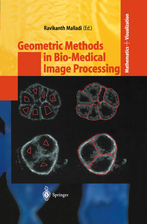
Geometric Methods in Bio-Medical Image Processing
Springer Berlin (Verlag)
9783540432166 (ISBN)
Itgivesmegreatpleasuretoeditthisbook. Thegenesisofthisbookgoes backtotheconferenceheldattheUniversityofBolognainJune1999,on collaborativeworkbetweentheUniversityofCaliforniaatBerkeleyandthe UniversityofBologna. Theoriginalideawastoinvitesomespeakersatthe conferencetosubmitarticlestothebook. Thescopeofthebookwaslater- hancedand,inthepresentform,itisacompilationofsomeoftherecentwork usinggeometricpartialdi?erentialequationsandthelevelsetmethodology inmedicalandbiomedicalimageanalysis. Thesynopsisofthebookisasfollows:Inthe?rstchapter,R. Malladi andJ. A. Sethianpointtotheoriginsoftheuseoflevelsetmethodsand geometricPDEsforsegmentation,andpresentfastmethodsforshapes- mentationinbothmedicalandbiomedicalimageapplications. InChapter 2,C. OrtizdeSolorzano,R. Malladi,andS. J. Lockettdescribeabodyof workthatwasdoneoverthepastcoupleofyearsattheLawrenceBerkeley NationalLaboratoryonapplicationsoflevelsetmethodsinthestudyand understandingofconfocalmicroscopeimagery. TheworkinChapter3byA. Sarti,C. Lamberti,andR. Malladiaddressestheproblemofunderstanding di?culttimevaryingechocardiographicimagery. Thisworkpresentsvarious levelsetmodelsthataredesignedto?tavarietyofimagingsituations,i. e. timevarying2D,3D,andtimevarying3D. InChapter4,L. VeseandT. F. Chanpresentasegmentationmodelwithoutedgesandalsoshowextensions totheMumford-Shahmodel. Thismodelisparticularlypowerfulincertain applicationswhencomparisonsbetweennormalandabnormalsubjectsis- quired. Next,inChapter5,A. EladandR. Kimmelusethefastmarching methodontriangulateddomaintobuildatechniquetounfoldthecortexand mapitontoasphere. Thistechniqueismotivatedinpartbynewadvances infMRIbasedneuroimaging. InChapter6,T. DeschampsandL. D. Cohen presentaminimalpathbasedmethodofgroupingconnectedcomponentsand showcleverapplicationsinvesseldetectionin3Dmedicaldata. Finally,in Chapter7,A. Sarti,K. Mikula,F. Sgallari,andC. Lamberti,describean- linearmodelfor?lteringtimevarying3Dmedicaldataandshowimpressive resultsinbothultrasoundandechoimages. IoweadebtofgratitudetoClaudioLambertiandAlessandroSartifor invitingmetoBologna,andlogisticalsupportfortheconference. Ithank thecontributingauthorsfortheirenthusiasmand?exibility,theSpringer mathematicseditorMartinPetersforhisoptimismandpatience,andJ. A. Sethianforhisunfailingsupport,goodhumor,andguidancethroughthe years. Berkeley,California R. Malladi October,2001 Contents 1 FastMethodsforShapeExtractioninMedicaland BiomedicalImaging. . . . . . . . . . . . . . . . . . . . . . . . . . . . . . . . . . . . . . . . . . . 1 R. Malladi,J. A. Sethian 1. 1Introduction. . . . . . . . . . . . . . . . . . . . . . . . . . . . . . . . . . . . . . . . . . . . . . . . 1 1. 2TheFastMarchingMethod. . . . . . . . . . . . . . . . . . . . . . . . . . . . . . . . . . . 3 1. 3ShapeRecoveryfromMedicalImages. . . . . . . . . . . . . . . . . . . . . . . . . . 6 1. 4Results. . . . . . . . . . . . . . . . . . . . . . . . . . . . . . . . . . . . . . . . . . . . . . . . . . . . . 10 References. . . . . . . . . . . . . . . . . . . . . . . . . . . . . . . . . . . . . . . . . . . . . . . . . . . . . 13 2 AGeometricModelforImageAnalysisinCytology. . . . . . . 19 C. OrtizdeSolorzano,R. Malladi,,S. J. Lockett 2. 1Introduction. . . . . . . . . . . . . . . . . . . . . . . . . . . . . . . . . . . . . . . . . . . . . . . . 19 2. 2GeometricModelforImageAnalysis. . . . . . . . . . . . . . . . . . . . . . . . . . . 20 2. 3SegmentationofNuclei. . . . . . . . . . . . . . . . . . . . . . . . . . . . . . . . . . . . . . . 22 2. 4SegmentationofNucleiandCellsUsingMembrane-RelatedProtein Markers. . . . . . . . . . . . . . . . . . . . . . . . . . . . . . . . . . . . . . . . . . . . . . . . . . . . 31 2. 5Conclusions. . . . . . . . . . . . . . . . . . . . . . . . . . . . . . . . . . . . . . . . . . . . . . . . . 37 References. . . . . . . . . . . . . . . . . . . . . . . . . . . . . . . . . . . . . . . . . . . . . . . . . . . . . 38 3 LevelSetModelsforAnalysisof2Dand3D EchocardiographicData. . . . . . . . . . . . . . . .
1 Fast Methods for Shape Extraction in Medical and Biomedical Imaging.- 1.1 Introduction.- 1.2 The Fast Marching Method.- 1.3 Shape Recovery from Medical Images.- 1.4 Results.- References.- 2 A Geometric Model for Image Analysis in Cytology.- 2.1 Introduction.- 2.2 Geometric Model for Image Analysis.- 2.3 Segmentation of Nuclei.- 2.4 Segmentation of Nuclei and Cells Using Membrane-Related Protein Markers.- 2.5 Conclusions.- References.- 3 Level Set Models for Analysis of 2D and 3D Echocardiographic Data.- 3.1 Introduction.- 3.2 The Geometric Evolution Equation.- 3.3 The Shock-Type Filtering.- 3.4 Shape Extraction.- 3.5 2D Echocardiography.- 3.6 2D + time Echocardiography.- 3.7 3D Echocardiography.- 3.8 3D + time Echocardiography.- 3.9 Conclusions.- References.- 4 Active Contour and Segmentation Models using Geometric PDE's for Medical Imaging.- 4.1 Introduction.- 4.2 Description of the Models.- 4.3 Applications to Bio-Medical Images.- 4.4 Concluding Remarks.- References.- 5 Spherical Flattening of the Cortex Surface.- 5.1 Introduction.- 5.2 Fast Marching Method on Triangulated Domains.- 5.3 Multi-Dimensional Scaling.- 5.4 Cortex Unfolding.- 5.5 Conclusions.- References.- 6 Grouping Connected Components using Minimal Path Techniques.- 6.1 Introduction.- 6.2 Minimal Paths in 2D and 3D.- 6.3 Finding Contours from a Set of Connected Components Rk.- 6.4 Finding a Set of Paths in a 3D Image.- 6.5 Conclusion.- References.- 7 Nonlinear Multiscale Analysis Models for Filtering of 3D + Time Biomedical Images.- 7.1 Introduction.- 7.2 Nonlinear Diffusion Equations for Processing of 2D and 3D Still*Images.- 7.3 Space-Time Filtering Nonlinear Diffusion Equations.- 7.4 Numerical Algorithm.- 7.5 Discussion on Numerical Experiments.- 7.6 Preconditioning and Solving of Linear Systems.-References.- Appendix. Color Plates.
From the reviews:
R. Malladi (ed.)
Geometric Methods in Bio-Medical Image Processing
"This is an excellent monograph on geometric methods in biomedical image processing. I strongly recommend this book to visualization experts in mathematics, computer science and bio-medical applications and to research students on above topics."-JOURNAL OF COMPUTATIONAL METHODS IN APPLIED MATHEMATICS
"This book is based on the conference held at the University of Bologna at June 1999. ... The book gives a good review on some of the traditional applications in medical imagery (CT, MR, Ultrasound). ... This is an excellent monograph on geometric methods in biomedical image processing. I strongly recommend this book to visualization experts in mathematics, computer science and bio-medical applications and to research students on the above topics." (T. E. Simos, Journal of Computational Methods in Sciences and Engineering, Vol. 3 (2), 2003)
| Erscheint lt. Verlag | 23.4.2002 |
|---|---|
| Reihe/Serie | Mathematics and Visualization |
| Zusatzinfo | VIII, 147 p. |
| Verlagsort | Berlin |
| Sprache | englisch |
| Maße | 155 x 235 mm |
| Gewicht | 424 g |
| Themenwelt | Informatik ► Software Entwicklung ► User Interfaces (HCI) |
| Mathematik / Informatik ► Mathematik ► Graphentheorie | |
| Mathematik / Informatik ► Mathematik ► Wahrscheinlichkeit / Kombinatorik | |
| Medizinische Fachgebiete ► Radiologie / Bildgebende Verfahren ► Radiologie | |
| Schlagworte | algorithm • Bildanalyse (EDV) • Biomedical Images • Calculus • Computed tomography (CT) • Computergestützte Bildgebende Verfahren (Medizin) • Cytometry • Diffusion • Echocardiography • filtering • Image Analysis • Image Processing • Level Sets • Life Sciences • Medical Imaging • Methodology • Protein • Ultrasound • Ultrasound Imagery |
| ISBN-13 | 9783540432166 / 9783540432166 |
| Zustand | Neuware |
| Informationen gemäß Produktsicherheitsverordnung (GPSR) | |
| Haben Sie eine Frage zum Produkt? |
aus dem Bereich


