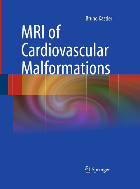
MRI of Cardiovascular Malformations
Springer Berlin (Verlag)
978-3-642-43838-7 (ISBN)
MRI is a non-invasive and non-ionizing imaging modality that is perfectly suited for the diagnosis and follow-up of both pediatric and adult congenital heart disease. It provides a large field of view and has the unique ability to depict complex cardiac and vascular anatomy and to measure cardiac function and flow within one examination. MRI is the ideal complement to echocardiography whenever the information provided by the latter is limited.This book has been conceived as a self-teaching manual that will assist qualified radiologists, cardiologists, and pediatricians, as well as those in training. It is richly illustrated with numerous images and drawings that cover all usual and most unusual anomalies. The principal author, Professor Bruno Kastler, is head of radiology at Besançon University Hospital, France and is board certified in both radiology and cardiology.
Professor Bruno Kastler completed his MS degree in Physiscs (1994- 1977) and his MD degree (1994- 1981) at the Université de la Méditerranée Marseille. France. He spent Two years in the Physiology Department of the University of Minnesota as post doctorate (1981-84) and his residency first in Minneapolis and then in Strasbourg at the University Hospital where he was Board certified in cardiology (1987) and Radiology (1988) at the Université Louis Pasteur. He was fellow in the Radiology department of University Hospital of Strasbourg (1988-94). He is currently Head of the department of diagnostic and interventional radiology at the University Hospital of Besançon (1994), Director of I4S Laboratory (Health Innovation Intervention, Imaging, and Engineering) University of Franche-Comté (1997) and Associate Professor University of Sherbrook Canada (1992). He is the Laureate of the first prize of the Victoires de la médecine in innovating techniques (Paris 2006) and Doctor Honoris Causae of the University of Sophia (2006). His fields of interest cover physical principals and technical aspects of MRI , cardiovascular disease and interventional radiology using CT-guidance in the treatment of pain. He is the author of books on each of these topics.
Patient Preparation: Magnetic Resonance Imaging Techniques.- Cardiovascular Anatomy and Atlas of MR Normal Anatomy.- MRI Sequential Segmental Analysis and Identification of Anomalies.- Anomalous Systemic and Pulmonary Venous Connections.- Aortic Arch Anomalies, Tracheal Compression and Stenosis.- Other Aortic Malforamtions.- Anomalies of the Right Ventricular Outflow Tract and Pulmonary Arteries: Complex Congenital Heart Disease.- Postoperative Evaluation.
From the reviews:
"The book is split into eight sections covering patient preparation and techniques ... . The text and drawings make even the more complex lesion understandable with clear and logical descriptions used throughout. ... it is reasonably priced and should act as a 'bench book' in any department that undertakes imaging in congenital heart disease. Although it appears that this book is aimed more at the specialist than general audience, it would also be a useful text for the incidental finding on a general CMR list." (Bobby Agrawal, RAD Magazine, October, 2011)| Erscheint lt. Verlag | 13.12.2014 |
|---|---|
| Zusatzinfo | XVII, 256 p. |
| Verlagsort | Berlin |
| Sprache | englisch |
| Maße | 193 x 260 mm |
| Gewicht | 581 g |
| Themenwelt | Medizinische Fachgebiete ► Innere Medizin ► Kardiologie / Angiologie |
| Medizin / Pharmazie ► Medizinische Fachgebiete ► Pädiatrie | |
| Medizinische Fachgebiete ► Radiologie / Bildgebende Verfahren ► Radiologie | |
| Schlagworte | Abnormalities • Cardiac Anomalies • Cardiovascular Magnetic Resonance • Congenital • Congenital Heart Disease |
| ISBN-10 | 3-642-43838-5 / 3642438385 |
| ISBN-13 | 978-3-642-43838-7 / 9783642438387 |
| Zustand | Neuware |
| Haben Sie eine Frage zum Produkt? |
aus dem Bereich


