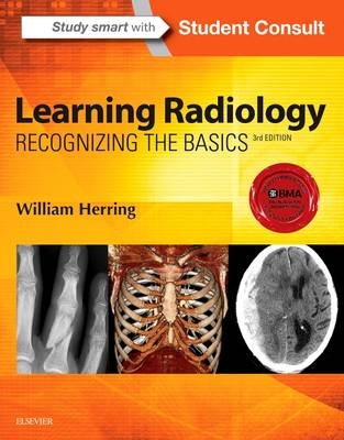
Learning Radiology
Saunders (Verlag)
978-0-323-32807-4 (ISBN)
- Titel erscheint in neuer Auflage
- Artikel merken
Identify a wide range of common and uncommon conditions based upon their imaging findings.
Arrive at diagnoses by following a pattern recognition approach, and logically overcome difficult diagnostic challenges with the aid of decision trees.
Quickly grasp the fundamentals you need to know through more than 700 images and an easy-to-use format and pedagogy, including: bolding of key points and icons designating special content; Diagnostic Pitfalls; Really, Really Important Points; Weblinks; and Take-Home Points.
Gauge your mastery of the material and build confidence with extra images, bonus content, interactive self-assessment exercises, and USMLE-style Q&A that provide effective chapter review and quick practice for your exams.
Apply the latest recommendations on patient safety, dose reduction and radiation protection
Benefit from the extensive knowledge and experience of esteemed author Dr. William Herring-a skilled radiology teacher and the host of his own specialty website, www.learningradiology.com.
Stay current in the latest advancements and developments with meticulous updates throughout including a new chapter on Pediatric Radiology as well as more than 60 new and updated photos, many highlighting newer imaging modalities.
Maximize your learning experience with interactive Student Consult extras videos/images of 3D images, functional imaging examinations, dynamic studies, and additional assessments.
Student Consult eBook version included with purchase. This enhanced eBook experience allows you to search all of the text, figures, references, and videos from the book on a variety of devices.
Chapter 1. Recognizing Anything
Chapter 2. Recognizing a Technically Adequate Chest Radiograph
Chapter 3. Recognizing Normal Pulmonary Anatomy
Chapter 4. Recognizing Normal Cardiac Anatomy
Chapter 5. Recognizing Airspace versus Interstitial Lung Disease
Chapter 6. Recognizing the Causes of an Opacified Hemithorax
Chapter 7. Recognizing Atelectasis
Chapter 8. Recognizing a Pleural Effusion
Chapter 9. Recognizing Pneumonia
Chapter 10. Recognizing Pneumothorax, Pneumomediastinum, Pneumopericardium, and Subcutaneous Emphysema
Chapter 11. Recognizing the Correct Placement of Lines and Tubes and Their Potential Complications: Critical Care Radiology
Chapter 12. Recognizing Diseases of the Chest
Chapter 13: Recognizing Adult Heart Disease
Chapter 14. Recognizing the Normal Abdomen: Conventional Radiology
Chapter 15. Recognizing the Normal Abdomen and Pelvis on Computed Tomography
Chapter 16. Recognizing Bowel Obstruction and Ileus
Chapter 17. Recognizing Extraluminal Gas in the Abdomen
Chapter 18. Recognizing Abnormal Calcifications and Their Causes
Chapter 19. Recognizing the Imaging Findings of Trauma
Chapter 20. Recognizing Gastrointestinal, Hepatic, and Urinary Tract Abnormalities
Chapter 21. Ultrasonography: Understanding the Principles and Recognizing Normal and Abnormal Findings
Chapter 22. Magnetic Resonance Imaging: Understanding the Principles and Recognizing the Basics
Daniel J. Kowal, MD
Chapter 23. Recognizing Abnormalities of Bone Density
Chapter 24. Recognizing Fractures and Dislocations
Chapter 25. Recognizing Joint Disease: An Approach to Arthritis
Chapter 26. Recognizing Some Common Causes of Neck and Back Pain
Chapter 27. Recognizing Some Common Causes of Intracranial Pathology
Chapter 28. Recognizing Pediatric Diseases
Appendix: What to Order When
Bibliography
Chapter 1 Quiz Answers
Online Content
| Zusatzinfo | Approx. 656 illustrations (15 in full color) |
|---|---|
| Verlagsort | Philadelphia |
| Sprache | englisch |
| Maße | 216 x 276 mm |
| Themenwelt | Medizinische Fachgebiete ► Radiologie / Bildgebende Verfahren ► Radiologie |
| ISBN-10 | 0-323-32807-5 / 0323328075 |
| ISBN-13 | 978-0-323-32807-4 / 9780323328074 |
| Zustand | Neuware |
| Informationen gemäß Produktsicherheitsverordnung (GPSR) | |
| Haben Sie eine Frage zum Produkt? |
aus dem Bereich


