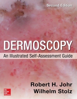
Dermoscopy: An Illustrated Self-Assessment Guide, 2/e
Seiten
2015
|
2nd edition
McGraw-Hill Professional (Verlag)
978-0-07-183434-6 (ISBN)
McGraw-Hill Professional (Verlag)
978-0-07-183434-6 (ISBN)
Dermoscopy: An Illustrated Self-Assessment Guide offers a simple, innovative, and highly visual approach to learning the general principles, terminology, and specific criteria of dermoscopy usage.
Publisher's Note: Products purchased from Third Party sellers are not guaranteed by the publisher for quality, authenticity, or access to any online entitlements included with the product.
Learn dermoscopy with this full-color, case-based self-assessment guide
Awarded First Prize in the Internal Medicine category of the British Medical Association Medical Book Awards!
With 436 clinical and dermoscopic images and 218 progressively more difficult cases commonly encountered in general dermatologic practice, Dermoscopy: An Illustrated Self-Assessment Guide offers a unique checklist methodology for learning how to use dermosocpy to diagnose benign and malignant pigmented and non-pigmented skin lesions.
Each high-quality, full-color clinical and dermoscopic image is presented with short history. Every case is followed by multiple-choice questions and three check boxes to test your knowledge of risk, diagnosis, and disposition. Turn the page, and the answers to the questions are provided in an easy-to-remember manner which includes the dermoscopic images being sown again. Circles, stars, boxes, and arrows appear in the image pointing out the important criteria of each case.
FEATURES:
Cases involving the scalp, face, nose, ears, trunk and extremities, palms, soles, nails, and genitalia – many new to this edition
The concepts of clinic-dermoscopic correlation, dermoscopic-pathologic correlation, and dermoscopic differential diagnosis are employed throughout
Each case includes a discussion of all of its salient features in a quick-read outline style and ends with a series of dermoscopic and/or clinical pearls based on the authors’ years of experience
Key dermoscopic principles are re-emphasized throughout the book to enhance your understanding and assimilation of the teaching points
Two new chapters on trichoscopy and dermoscopy in general medicine
Updated material on pediatric melanoma, desmoplastic melanoma, Merkel cell carcinoma, invasive squamous cell carcinoma, and nevi and melanoma associated with decorative tattoos
Robert H. Johr, MD is Clinical Professor of Dermatology, Associate Clinical Professor of Pediatrics, Pigmented Lesion Clinic, University of Miami School of Medicine. Wilhelm Stolz, MD is Director, Clinic of Dermatology, Allergy, and Environmental Medicine, Hospital Munchen Schwabing; Professor of Dermatology, Faculty of Medicine, Ludwig-Maximilians-Universitat.
1. Dermoscopy from A to Z
2. Scalp, Face, Nose, and Ears
3. Trunk and Extremities
4. Palms, Soles, Nails
5. Genitalia
| Erscheint lt. Verlag | 16.10.2015 |
|---|---|
| Zusatzinfo | 0 Illustrations |
| Sprache | englisch |
| Maße | 218 x 274 mm |
| Gewicht | 1283 g |
| Themenwelt | Medizin / Pharmazie ► Medizinische Fachgebiete ► Dermatologie |
| ISBN-10 | 0-07-183434-6 / 0071834346 |
| ISBN-13 | 978-0-07-183434-6 / 9780071834346 |
| Zustand | Neuware |
| Haben Sie eine Frage zum Produkt? |
Mehr entdecken
aus dem Bereich
aus dem Bereich
Differentialdiagnostik und Therapie bei Kindern und Jugendlichen
Buch (2022)
Thieme (Verlag)
CHF 191,00
Allergene - Diagnostik - Therapie
Buch (2022)
Thieme (Verlag)
CHF 249,00
richtig verschreiben – individuell therapieren
Buch (2023)
Thieme (Verlag)
CHF 99,40


