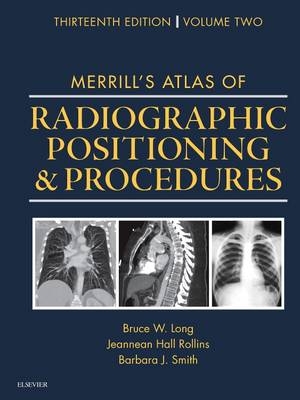
Merrill's Atlas of Radiographic Positioning and Procedures
Volume 2
Seiten
2015
|
13th Revised edition
Mosby (Verlag)
978-0-323-26343-6 (ISBN)
Mosby (Verlag)
978-0-323-26343-6 (ISBN)
- Titel erscheint in neuer Auflage
- Artikel merken
Zu diesem Artikel existiert eine Nachauflage
With more than 400 projections presented, Merrill's Atlas of Radiographic Positioning and Procedures remains the gold standard of radiographic positioning texts. Authors Eugene Frank, Bruce Long, and Barbara Smith have designed this comprehensive resource to be both an excellent textbook and also a superb clinical reference for practicing radiographers and physicians. You'll learn how to properly position the patient so that the resulting radiograph provides the information needed to reach an accurate diagnosis. Complete information is included for the most common projections, as well as for those less commonly requested.
Comprehensive, full-color coverage of anatomy and positioning makes Merrill's Atlas the most in-depth text and reference available for radiography students and practitioners.
Frequently performed projections are identified with a special icon to help you focus on what you need to know as an entry-level radiographer.
Numerous CT and MRI images enhance your comprehension of cross-sectional anatomy and help you prepare for the Registry examination.
UNIQUE! Collimation sizes and other key information are provided for each relevant projection.
Bulleted lists provide clear instructions on how to correctly position the patient and body part when performing procedures.
Summary tables provide quick access to projection overviews, guides to anatomy, pathology tables for bone groups and body systems, and exposure technique charts.
NEW! Coverage of the latest advances in digital imaging also includes more digital radiographs with greater contrast resolution of pertinent anatomy.
NEW positioning photos show current digital imaging equipment and technology.
UPDATED coverage addresses contrast arthrography procedures, trauma radiography practices, plus current patient preparation, contrast media used, and the influence of digital technologies.
UPDATED Mammography chapter reflects the evolution to digital mammography, as well as innovations in breast biopsy procedures.
VOLUME 2
11. Long Bone Measurement
12. Contrast Arthrography
13. Trauma Radiography
14. Mouth and Salivary Glands
15. Anterior Part of Neck
16. Abdomen
17. Digestive System: Alimentary Canal
18. Urinary System and Venipuncture
19. Reproductive System
20. Skull, Facial Bones, and Paranasal Sinuses
21. Mammography
| Zusatzinfo | Approx. 1350 illustrations (580 in full color) |
|---|---|
| Verlagsort | St Louis |
| Sprache | englisch |
| Maße | 306 x 234 mm |
| Themenwelt | Medizin / Pharmazie ► Gesundheitsfachberufe ► MTA - Radiologie |
| Medizin / Pharmazie ► Medizinische Fachgebiete ► Radiologie / Bildgebende Verfahren | |
| ISBN-10 | 0-323-26343-7 / 0323263437 |
| ISBN-13 | 978-0-323-26343-6 / 9780323263436 |
| Zustand | Neuware |
| Haben Sie eine Frage zum Produkt? |
Mehr entdecken
aus dem Bereich
aus dem Bereich
Buch | Softcover (2024)
Studia Universitätsverlag Innsbruck
CHF 67,20
Buch | Hardcover (2024)
Mosby (Verlag)
CHF 319,95


