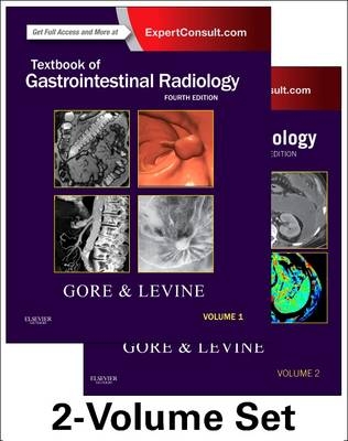
Textbook of Gastrointestinal Radiology, 2-Volume Set
W B Saunders Co Ltd (Verlag)
978-1-4557-5117-4 (ISBN)
- Titel erscheint in neuer Auflage
- Artikel merken
Characterize abdominal masses and adenopathy with the aid of diffusion-weighted MR imaging.
See how gastrointestinal conditions present with more than 2,500 multi-modality, high-quality digital images that mirror the findings you're likely to encounter in practice.
Make optimal use of the latest abdominal and gastrointestinal imaging techniques with new chapters on diffusion weighted MRI, perfusion MDCT and MRI, CT colonography, CT enterography and MR enterography-sophisticated cross-sectional imaging techniques that have dramatically improved the utility of CT and MR for detecting a host of pathologic conditions in the gastrointestinal tract.
Expert guidance is right at your fingertips. Now optimized for use on mobile devices, this edition is perfect as an on-the-go resource for all abdominal imaging needs.
Effectively apply MR and CT perfusion, diffusion weighted imaging, PET/CT and PET/MR in evaluating tumor response to therapy.
VOLUME 1 SECTION I: GENERAL RADIOLOGIC PRINCIPLES
1. Imaging Contrast Agents and Pharmacoradiology
2. Barium Studies: Single and Double
3. Pictorial Glossary of Double-Contrast Radiology
4. Ultrasound of the Hollow Viscera
5. Multidetector Computed Tomography of the Gastrointestinal Tract: Principles of Interpretation
6. Magnetic Resonance Imaging of the Hollow Viscera
7. Positron Emission Tomography and Computed Tomography of the Hollow Viscera
8. Angiography and Interventional Radiology of the Hollow Viscera
9. Abdominal Computed Tomography Angiography
10. Magnetic Resonance Angiography of the Mesenteric Vasculature
SECTION II: ABDOMINAL PLAIN IMAGES
10. Abdomen: Normal Anatomy and Examination Techniques
11. Gas and Soft Tissue Abnormalities
12. Abnormal Calcifications
SECTION III: PHARYNX
14. Pharynx: Normal Anatomy and Examination Techniques
15. Abnormalities of Pharyngeal Function
16. Structural Abnormalities of the Pharynx
SECTION IV: ESOPHAGUS
17. Barium Studies of the Upper Gastrointestinal Tract
18. Motility Disorders of the Esophagus
19. Gastroesophageal Reflux Disease
20. Infectious Esophagitis
21. Other Esophagitides
22. Benign Tumors of the Esophagus
23. Carcinoma of the Esophagus
24. Other Malignant Tumors of the Esophagus
25. Miscellaneous Abnormalities of the Esophagus
26. Abnormalities of the Gastroesophageal Junction
27. Postoperative Esophagus
28. Esophagus: Differential Diagnosis
SECTION V: STOMACH AND DUODENUM
29. Peptic Ulcers
30. Inflammatory Conditions of the Stomach and Duodenum
31. Benign Tumors of the Stomach and Duodenum
32. Carcinoma of the Stomach and Duodenum
33. Other Malignant Tumors of the Stomach and Duodenum
34. Miscellaneous Abnormalities of the Stomach and Duodenum
35. Postoperative Stomach and Duodenum
36. Stomach and Duodenum: Differential Diagnosis
SECTION VI: SMALL BOWEL
37. Barium Examinations of the Small Intestine
38. Computed Tomographic Enterography
39. Computed Tomography Enteroclysis
40. Magnetic Resonance Enterography
41. Crohn's Disease of the Small Bowel
42. Inflammatory Disorders of the Small Bowel Other Than Crohn's Disease
43. Malabsorption
44. Benign Tumors of the Small Bowel
45. Malignant Tumors of the Small Bowel
46. Small Bowel Obstruction
47. Vascular Disorders of the Small Intestine
48. Postoperative Small Bowel
49. Miscellaneous Abnormalities of the Small Bowel
50. Small Intestine: Differential Diagnosis
SECTION VII: COLON
51. Barium Studies of the Colon
52. Functional Imaging of Anorectal and Pelvic Floor Dysfunction
53. Computed Tomography Colonography
54. Magnetic Resonance Colonography
55. Diverticular Disease of the Colon
56. Diseases of the Appendix
57. Ulcerative and Granulomatous Colitis: Idiopathic Inflammatory Bowel Disease
58. Other Inflammatory Conditions of the Colon
59. Polyps and Colon Cancer
60. Other Tumors of the Colon
61. Polyposis Syndromes
62. Miscellaneous Abnormalities of the Colon
63. Postoperative Colon
64. Colon: Differential Diagnosis
VOLUME II SECTION VIII: General Radiologic Principles for Imaging and Intervention of the Solid Viscera
65. Computed Tomography of the Solid Abdominal Organs
66. Ultrasound Examination of the Solid Abdominal Organs
67. Magnetic Resonance Imaging of the Solid Parenchymal Organs
68. Positron Emission Tomography/Computed Tomography of the Solid Parenchymal Organs
69. Diffusion-Weighted Imaging of the Abdomen
70. Perfusion Computed Tomography and Magnetic Resonance Imaging in the Abdomen and Pelvis
71. Techniques of Percutaneous Tissue Acquisition
72. Abdominal Abcess
SECTION IX: GALLBLADDER AND BILIARY TRACT
73. Gallbladder and Biliary Tract: Normal Anatomy and Examination Techniques
74. Endoscopic Retrograde Cholangiopancreatography
75. Magnetic Resonance Cholangiopancreatography
76. Anomalies and Anatomic Variants of the Gallbladder and Biliary Tract
77. Cholelithiasis, Cholecystitis, Choledocholithasis, and Hyperplastic Cholecystoses
78. Interventional Radiology of the Gallbladder and Biliary Tract
79. Neoplasms of the Gallbladder and Biliary Tract
80. Inflammatory Disorders of the Biliary Tract
81. Postsurgical and Traumatic Lesions of the Biliary Tract
82. Gallbladder and Biliary Tract: Differential Diagnosis
SECTION X: LIVER
83. Liver: Normal Anatomy and Examination Techniques
84. Interventional Radiology of the Liver
85. Anomalies and Anatomic Variants of the Liver
86. Benign Tumors of the Liver
87. Malignant Tumors of the Liver
88. Focal Hepatic Infections
89. Diffuse Liver Disease
90. Vascular Disorders of the Liver and Splanchnic Circulation
91. Hepatic Trauma, Surgery, and Liver-Directed Therapy
92. Liver Transplantation Imaging
93. Liver: Differential Diagnosis
SECTION XI: PANCREAS
94. Pancreas: Normal Anatomy and Examination Techniques
95. Interventional Radiology of the Pancreas
96. Anomalies and Anatomic Variants of the Pancreas
97. Pancreatitis
98. Pancreatic Neoplasms
99. Pancreatic Trauma and Surgery
100. Pancreatic Transplantation Imaging
101. Pancreas: Differential Diagnosis
SECTION XII: SPLEEN
102. Spleen: Normal Anatomy and Examination Techniques
103. Angiography and Interventional Radiology of the Spleen
104. Anomalies and Anatomic Variants of the Spleen
105. Benign and Malignant Lesions of the Spleen
106. Splenic Trauma and Surgey
107. Spleen: Differential Diagnosis
SECTION XIII: PERITONEAL CAVITY
108. Anatomy and Imaging of the Peritoneum and Retroperitoneum
109. Pathways of Abdominal and Pelvic Disease Spread
110. Ascites and Peritoneal Fluid Collections
111. Mesenteric and Omental Lesions
112. Hernias and Abdominal Wall Pathology
SECTION XIV: PEDIATRIC DISEASE
113. Applied Embryology of the Gastrointestinal Tract
114. Neonatal Gastrointestinal Radiology
115. Diseases of the Pediatric Esophagus
116. Diseases of the Pediatric Stomach and Duodenum
117. Diseases of the Pediatric Small Bowel
118. Diseases of the Pediatric Colon
119. Diseases of the Pediatric Gallblader and Biliary Tract
120. Diseases of the Pediatric Liver
121. Diseases of the Pediatric Pancreas
122. Diseases of the Pediatric Spleen
123. Diseases of the Pediatric Abdominal Wall, Peritoneum, and Mesentery
SECTION XV: COMMON CLINICAL PROBLEMS
124. The Acute Abdomen
125. Gastrointestinal Hemorrhage
126. Abdominal Trauma
127. Monitoring Gastrointestinal Tumor Response to Therapy
| Erscheint lt. Verlag | 24.6.2021 |
|---|---|
| Zusatzinfo | Approx. 2050 illustrations (650 in full color); Illustrations, unspecified |
| Verlagsort | London |
| Sprache | englisch |
| Maße | 216 x 286 mm |
| Gewicht | 7290 g |
| Themenwelt | Medizinische Fachgebiete ► Innere Medizin ► Gastroenterologie |
| Medizinische Fachgebiete ► Radiologie / Bildgebende Verfahren ► Radiologie | |
| ISBN-10 | 1-4557-5117-0 / 1455751170 |
| ISBN-13 | 978-1-4557-5117-4 / 9781455751174 |
| Zustand | Neuware |
| Haben Sie eine Frage zum Produkt? |
aus dem Bereich


