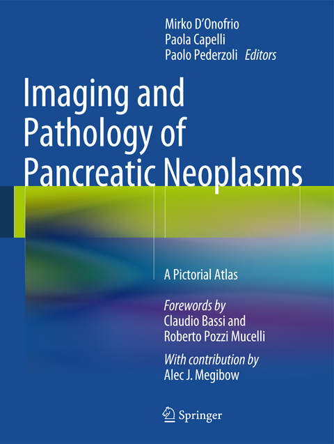
Imaging and Pathology of Pancreatic Neoplasms
A Pictorial Atlas
Seiten
2014
|
2015 ed.
Springer Verlag
978-88-470-5677-0 (ISBN)
Springer Verlag
978-88-470-5677-0 (ISBN)
Zu diesem Artikel existiert eine Nachauflage
Imaging and Pathology of Pancreatic Neoplasms
Although interest in pancreatic pathology is very high in the radiological and gastroenterological communities, it is still the case that less is known about pathology of the pancreas than about liver pathology, for example. Diagnosis depends on the structure of the pancreatic lesion, which can be directly visualized on US, CT or MR images. This atlas, which encompasses both the imaging and the pathology of pancreatic neoplasms, will therefore be invaluable in enabling radiologists and sonographers to understand the underlying pathology and in allowing pancreatic pathologists to understand the imaging translation.
The emphasis in the atlas is very much on the pathological and imaging appearances, with most of the text concentrated at the beginning of the chapters. A comprehensive overview is provided of typical and atypical presentations and diverse aspects of common and uncommon pancreatic neoplasms, including ductal adenocarcinoma, neuroendocrine neoplasms, intraductal papillary mucinous neoplasms, cystic neoplasms, metastases and lymphoma.
Ductal Adenocarcinoma.- Neuroendocrine Neoplasm.- Intraductal Neoplasms.- Serous Neoplasms.- Mucinous Neoplasms.- Solid-Pseudopapillary Neoplasms.- Secondary Tumors and Lymphoma.- Rare Neoplasms.- Driving Questions and Flow Charts Imaging.- Stigmate Review and Interpretation.
| Co-Autor | Alec J. Megibow |
|---|---|
| Vorwort | Claudio Bassi |
| Zusatzinfo | 213 Illustrations, color; 114 Illustrations, black and white; XIII, 432 p. 327 illus., 213 illus. in color. |
| Verlagsort | Milan |
| Sprache | englisch |
| Maße | 210 x 279 mm |
| Gewicht | 1469 g |
| Themenwelt | Medizin / Pharmazie ► Medizinische Fachgebiete ► Chirurgie |
| Medizinische Fachgebiete ► Innere Medizin ► Gastroenterologie | |
| Medizin / Pharmazie ► Medizinische Fachgebiete ► Onkologie | |
| Medizinische Fachgebiete ► Radiologie / Bildgebende Verfahren ► Radiologie | |
| Studium ► 2. Studienabschnitt (Klinik) ► Pathologie | |
| Schlagworte | Common and uncommon tumors • Ductal adenocarcinoma • Rare neoplasms • Secondary tumors and lymphoma • Solid Pseudopapillary tumors |
| ISBN-10 | 88-470-5677-2 / 8847056772 |
| ISBN-13 | 978-88-470-5677-0 / 9788847056770 |
| Zustand | Neuware |
| Haben Sie eine Frage zum Produkt? |
Mehr entdecken
aus dem Bereich
aus dem Bereich
Buch | Softcover (2024)
Urban & Fischer in Elsevier (Verlag)
CHF 76,00


