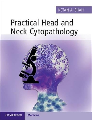
Practical Head and Neck Cytopathology with Online Static Resource
Cambridge University Press
978-1-107-44323-5 (ISBN)
Practical Head and Neck Cytopathology provides an engaging and holistic approach to the cytologic diagnosis of head and neck masses, covering both the technique of needle aspiration and its microscopic interpretation. A suitable sample is critical for arriving at the diagnosis and this book provides organ-specific tips on the FNA technique. Presence of different organs and structures in the head and neck region, as well as morphologic overlap amongst disparate entities, adds to the complexity of cytologic diagnosis. This book presents an easy-to-follow, stepwise approach to diagnosis, using concise bulleted text to highlight key features. Common and infrequently encountered mimics are included and interpretative challenges are discussed, facilitating a robust diagnosis. Numerous high-quality photomicrographs aid recognition of both common and rare features. A valuable resource for trainee and practising pathologists alike, this book is based on universally-used techniques and routinely prepared material, which are relevant to cytopathologists across the globe.
Ketan A. Shah is Consultant Pathologist, John Radcliffe Hospital, Oxford University Hospitals NHS Trust, Oxford, UK.
Preface; Acknowledgement; 1. Aspiration technique and stains; 2. Lymph nodes; 2.1. Introduction; 2.2. Reactive lymph node hyperplasia; 2.3. Granulomatous inflammation; 2.4. Lymphoma; 2.5. Metastatic disease; 2.6. Miscellaneous lymph node conditions; 2.7. Approach to lymph node cytology; 3. Salivary glands; 3.1. Introduction and cytology of normal salivary glands; 3.2. Proposed reporting system of salivary gland aspirates; 3.3. Inflammatory salivary gland lesions; 3.4. Cystic salivary gland lesions; 3.5. Benign salivary gland neoplasms; 3.6. Basal cell neoplasms; 3.7. Oncocytic lesions; 3.8. Malignant salivary gland neoplasms; 3.9. Approach to salivary gland cytology; 3.10. Role of repeat FNA; 4. Thyroid; 4.1. Introduction; 4.2. Colloid goiter; 4.3. Thyroiditis; 4.4. Papillary carcinoma; 4.5. Follicular neoplasms; 4.6. Medullary carcinoma; 4.7. Poorly differentiated carcinoma; 4.8. Undifferentiated (anaplastic) carcinoma; 4.9. Hyalinizing trabecular tumour; 4.10. Lymphoma; 4.11. Approach to thyroid cytology; 5. Cystic neck lesions; 5.1. Introduction; 5.2. Branchial cyst; 5.3. Thyroglossal cyst; 5.4. Lymphangioma; 5.5. Seroma and lymphocele; 6. Other neck lesions; 6.1. Introduction; 6.2. Abscess; 6.3. Cervicofacial actinomycosis; 6.4. Parathyroid lesions; 6.5. Suture granuloma; 6.6. Lipoma; 6.7. Spindle cell lipoma; 6.8. Nodular fasciitis; 6.9. Paraganglioma; 6.10. Pilomatricoma; 6.11. Schwannoma; Index.
| Zusatzinfo | 6 Tables, black and white; 674 Halftones, color; 1 Line drawings, color |
|---|---|
| Verlagsort | Cambridge |
| Sprache | englisch |
| Maße | 195 x 255 mm |
| Gewicht | 970 g |
| Themenwelt | Studium ► 2. Studienabschnitt (Klinik) ► Pathologie |
| ISBN-10 | 1-107-44323-7 / 1107443237 |
| ISBN-13 | 978-1-107-44323-5 / 9781107443235 |
| Zustand | Neuware |
| Haben Sie eine Frage zum Produkt? |
aus dem Bereich

