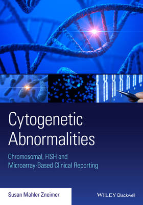
Cytogenetic Abnormalities
Wiley-Blackwell (Verlag)
978-1-118-91249-2 (ISBN)
Cytogenetics is the study of the structure and function of chromosomes in relation to phenotypic expression.Chromosomal abnormalities underlie the development of a wide variety of diseases and disorders ranging from Down syndrome to cancer, and are of widespread interest in both basic and clinical research.
Cytogenetic Abnormalities: Chromosomal, FISH, and Microarray-Based Clinical Reporting is a practical guide that describes cytogenetic abnormalities, their clinical implications and how best to report and communicate laboratory findings in research and clinical settings. The text first examines chromosomal, FISH, and microarray-based analyses in constitutional disorders. Using these same methodologies, the book's focus shifts to acquired abnormalities in cancers. Both sections provide illustrative examples of cytogenetic abnormalities and how to communicate these findings in standardized laboratory reports.
Providing both a wealth of cytogenetic information, as well as practical guidance on how best to communicate findings to fellow research and medical professionals, Cytogenetic Abnormalities will be an essential resource for cytogeneticists, laboratory personnel, clinicians, research scientists, and students in the field.
A guide to interpreting and reporting cytogenetic laboratory results involved in constitutional disorders and cancers
Guides the reader on implementing the International System for Human Cytogenetic Nomenclature in written reports
Provides information to allow scientists and medical professionals to fully understand and communicate cytogenetic abnormalities
Describes a wide array of cytogenetic abnormalities observed in the laboratory
Divided into user-friendly sections devoted to methodologies and implications of specific diseases
Susan Mahler Zneimer Ph.D., FACMGG is a clinical cytogeneticist and is the Scientific Director and CEO of MOSYS Consulting and Adjunct Professor at Moorpark College in Moorpark, California.
Preface xiv
Acknowledgments xv
About the companion website xvi
Introduction 1
Part 1: Constitutional Analyses 5
Section 1: Chromosome Analysis 7
1 Components of a standard cytogenetics report, normal results and culture failures 9
1.1 Components of a standard cytogenetics report 9
1.2 Prenatal normal results 17
1.3 Neonatal normal results 22
1.4 Normal variants in the population 23
1.5 Disclaimers and recommendations 29
1.6 Culture failures 30
1.7 Contamination 32
2 Mosaicism 35
2.1 Normal results with 30–50 cells examined 37
2.2 Normal and abnormal cell lines 37
2.3 Two or more abnormal cell lines 39
3 Autosomal trisomies – prenatal and livebirths 41
3.1 Introduction 41
3.2 Trisomy 21 – Down syndrome 42
3.3 Mosaic trisomy 21 – mosaic Down syndrome 43
3.4 Trisomy 13 – Patau syndrome 44
3.5 Trisomy 18 – Edwards syndrome 45
3.6 Trisomy 8 – mosaic 46
3.7 Trisomy 9 – mosaic 47
3.8 Trisomy 20 – mosaic, prenatal 47
3.9 Trisomy 22 – mosaic, prenatal 48
4 Translocations 51
4.1 Reciprocal (balanced) translocations 51
4.2 Robertsonian translocations 58
5 Inversions and recombinant chromosomes 67
5.1 Risks of spontaneous abortions and liveborn abnormal offspring 67
5.2 Pericentric inversions and their recombinants 67
5.3 Paracentric inversions and their recombinants 71
6 Visible deletions, duplications and insertions 75
6.1 Definitions 75
6.2 Visible duplications 79
6.3 Balanced insertions 80
7 Unidentifiable marker chromosomes, derivative chromosomes, chromosomes with additional material and rings 85
7.1 Marker chromosomes 85
7.2 Derivative chromosomes 87
7.3 Chromosomes with additional material 90
7.4 Ring chromosomes 91
7.5 Homogenously staining regions 94
8 Isochromosomes, dicentric chromosomes and pseudodicentric chromosomes 97
8.1 Isochromosomes/dicentric chromosomes 97
8.2 Pseudodicentric chromosomes 104
9 Composite karyotypes and other complex rearrangements 107
9.1 Composite karyotypes 107
9.2 Complex rearrangements 109
10 Sex chromosome abnormalities 115
10.1 X chromosome aneuploidies – female phenotypes 115
10.2 X and Y chromosome aneuploidies – male phenotypes 120
10.3 X chromosome structural abnormalities 122
10.4 Y chromosome structural abnormalities 128
10.5 46,XX males and 46,XY females 132
10.6 X chromosome translocations 135
11 Fetal demises/spontaneous abortions 143
11.1 Aneuploid rate 143
11.2 Confined placental mosaicism 144
11.3 Hydatidiform moles 145
11.4 Monosomy X in a fetus 146
11.5 Trisomies in a fetus 146
11.6 Double trisomy 149
11.7 Triploidy 150
11.8 Tetraploidy 151
12 Uniparental disomy 155
12.1 Uniparental disomy of chromosome 14 157
12.2 Uniparental disomy of chromosome 15 158
12.3 Uniparental disomy of chromosome 11p15 159
Section 2: Fluorescence In Situ Hybridization (FISH) Analysis 161
13 Metaphase analysis 163
13.1 Introduction 163
13.2 Reporting normal results 164
13.3 Common disclaimers 166
13.4 Microdeletions 167
13.5 Microduplications 190
13.6 Fluorescence in situ hybridization for chromosome identification 198
13.7 Subtelomere fluorescence in situ hybridization analysis 200
14 Interphase analysis 205
14.1 Introduction 205
14.2 Example report of interphase analysis 206
14.3 Common disclaimers 207
14.4 Reporting normal results 208
14.5 Abnormal prenatal/neonatal results 211
14.6 Abnormal product of conception FISH abnormalities 218
14.7 Molar pregnancies 222
14.8 Preimplantation genetic diagnosis 222
15 Integrated chromosome and FISH analyses 231
15.1 ISCN rules and reporting normal results by chromosomes and FISH 232
15.2 ISCN rules and reporting abnormal chromosomes and FISH 233
15.3 ISCN rules and reporting of chromosomes and subtelomere FISH 237
Section 3: Chromosomal Microarray Analysis (CMA) 243
16 Bacterial artificial chromosome, oligoarray and single nucleotide polymorphism array methodologies for analysis 245
16.1 Introduction 245
16.2 Clinical utility of chromosomal microarray analysis 250
16.3 Guidelines for classification states 251
16.4 ISCN rules and reporting of normal results 252
16.5 Comments, disclaimers and recommendations 253
17 Microarray abnormal results 257
17.1 Reporting of abnormal results 257
17.2 Loss or gain of a single chromosome 258
17.3 Loss or gain of a whole chromosome complement 262
17.4 Microdeletions 263
17.5 Microduplications 265
17.6 Derivative chromosomes 267
17.7 Variants of unknown significance 269
17.8 Uniparental disomy/loss of heterozygosity/regions of homozygosity 269
17.9 Mosaicism 271
17.10 Common comments in abnormal reports 273
17.11 Microarrays with concurrent FISH studies and/or chromosome studies 274
17.12 Microarrays with concurrent parental studies 274
17.13 Preimplantation genetic diagnosis testing 275
17.14 Non-invasive prenatal testing 276
18 Pathogenic chromosomal microarray copy number changes by chromosome order 285
18.1 Chromosome 1 285
18.2 Chromosome 2 287
18.3 Chromosome 3 289
18.4 Chromosome 4 290
18.5 Chromosome 5 291
18.6 Chromosome 7 292
18.7 Chromosome 8 293
18.8 Chromosome 14 294
18.9 Chromosome 15 294
18.10 Chromosome 16 296
18.11 Chromosome 17 298
18.12 Chromosome 19 301
18.13 Chromosome 22 302
18.14 Chromosome X 306
19 Integrated reports with cytogenetics, FISH and microarrays 315
19.1 Reporting of a deletion 315
19.2 Reporting of a supernumerary chromosome 316
19.3 Reporting of an unbalanced translocation – deletion/duplication 318
19.4 Reporting of multiple abnormal cell lines 322
Part 2: Acquired Abnormalities in Hematological and Tumor Malignancies 325
Section 1: Chromosome Analysis 327
20 Introduction 329
20.1 Description of World Health Organization classification for hematological malignancies 332
20.2 Description of different tumor types with significant cytogenetic abnormalities 332
20.3 Set-up and analysis of specific cultures for optimal results 333
20.4 Nomenclature rules for normal and simple abnormal results 336
20.5 Common report comments for hematological malignancies 338
21 Results with constitutional or other non-neoplastic abnormalities 347
21.1 Possible constitutional abnormalities observed 347
21.2 Age-related abnormalities 349
21.3 Non-clonal aberrations 351
21.4 No growth and poor growth 354
22 Cytogenetic abnormalities in myeloid disorders 357
22.1 Introduction to myeloid disorders 357
22.2 Individual myeloid abnormalities by chromosome order 360
23 Cytogenetic abnormalities in lymphoid disorders 395
23.1 Introduction to lymphoid disorders 395
23.2 Hyperdiploidy and hypodiploidy 396
23.3 Individual lymphoid abnormalities by chromosome order 398
24 Common biphenotypic abnormalities and secondary changes 423
24.1 Translocation (4;11)(q21;q23) 423
24.2 Del(9q) 424
24.3 Translocation (11;19)(q23;p13.3) 424
24.4 Del(12)(p11.2p13) 425
24.5 Trisomy 15 425
24.6 i(17q) 426
25 Reporting complex abnormalities and multiple cell lines 429
25.1 Stemline and sideline abnormalities 430
25.2 Unrelated abnormal clones 434
25.3 Composite karyotypes 435
25.4 Double minute chromosomes 436
25.5 Modal ploidy numbers 438
25.6 Multiple abnormal cell lines indicative of clonal evolution 440
26 Breakage disorders 445
26.1 Ataxia telangiectasia 445
26.2 Bloom syndrome 446
26.3 Fanconi anemia 447
26.4 Nijmegen syndrome 448
27 Cytogenetic abnormalities in solid tumors 451
27.1 Clear cell sarcoma 451
27.2 Chondrosarcoma 452
27.3 Ewing sarcoma 453
27.4 Liposarcoma 453
27.5 Neuroblastoma 454
27.6 Rhabdomyosarcoma 455
27.7 Synovial sarcoma 456
27.8 Wilms tumor 456
Section 2: Fluorescence In Situ Hybridization (FISH) Analysis 459
28 Introduction to FISH analysis for hematological disorders and solid tumors 461
28.1 General results 462
28.2 Bone marrow transplantation results 468
29 Recurrent FISH abnormalities in myeloid disorders 471
29.1 Individual abnormalities in myeloid disorders by chromosome order 471
29.2 Biphenotypic and therapy-related abnormalities 491
29.3 Panels of probes 492
30 Recurrent FISH abnormalities in lymphoid disorders 499
30.1 Individual abnormalities in lymphoid disorders by chromosome order 499
30.2 Panels of probes 520
31 Integrated reports with cytogenetics and FISH in hematological malignancies 531
31.1 Translocation (9;22) with BCR/ABL1 FISH analysis 531
31.2 Monosomy 7 with a marker chromosome and chromosome 7 FISH analysis 532
31.3 Complex abnormalities with the MDS FISH panel 532
31.4 Complex abnormalities with ALL FISH panel 533
31.5 Complex abnormalities with MM FISH panel 535
31.6 Complex abnormalities with AML FISH panel 536
31.7 Complex abnormalities with AML FISH panel in therapy-related disease 537
32 Recurrent FISH abnormalities in solid tumors using paraffin-embedded tissue 541
32.1 Ewing sarcoma 544
32.2 Liposarcoma 545
32.3 Neuroblastoma 546
32.4 Non-small cell lung cancer 547
32.5 Oligodendroglioma 552
32.6 Rhabdomyosarcoma 554
32.7 Synovial sarcoma 554
33 Breast cancer – HER2 FISH analysis 559
33.1 Common report comments 560
33.2 Example HER2 reports 561
33.3 Genetic heterogeneity 563
34 Bladder cancer FISH analysis 569
34.1 Common report comments 570
34.2 Example reports 570
Section 3: Chromosomal Microarray Analysis (CMA) 577
35 Chromosomal microarray analysis for hematological disorders 579
35.1 Introduction 579
35.2 Categories of abnormalities 580
35.3 Complex abnormalities throughout the genome, chromothripsis and homozygosity 581
35.4 Normal results and disclaimers 582
35.5 Example abnormal results in hematological malignancies 583
36 Chromosomal microarrays for tumors 595
36.1 Introduction and disclaimers 595
36.2 Breast cancer 596
36.3 Lung cancer 604
36.4 Colon cancer 606
36.5 Prostate cancer 607
36.6 Unspecified tumor present 607
37 Integrated reports with chromosomes, FISH and microarrays 611
37.1 Homozygous deletion of 9p21 identified by FISH and CMA 611
37.2 Identifying marker chromosomes by chromosome analysis, FISH and CMA 612
37.3 Unbalanced translocation identification by chromosomes, FISH and CMA 614
Appendix 1: Example assay-specific reagent (ASR) FISH validation plan for constitutional disorders and hematological malignancies on fresh tissue 617
Glossary 623
Index 641
| Verlagsort | Hoboken |
|---|---|
| Sprache | englisch |
| Maße | 178 x 254 mm |
| Gewicht | 1225 g |
| Themenwelt | Medizin / Pharmazie ► Medizinische Fachgebiete |
| Naturwissenschaften ► Biologie ► Zellbiologie | |
| ISBN-10 | 1-118-91249-7 / 1118912497 |
| ISBN-13 | 978-1-118-91249-2 / 9781118912492 |
| Zustand | Neuware |
| Informationen gemäß Produktsicherheitsverordnung (GPSR) | |
| Haben Sie eine Frage zum Produkt? |
aus dem Bereich


