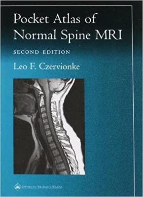
Pocket Atlas of Spinal MRI
Seiten
2000
|
2nd edition
Lippincott Williams and Wilkins (Verlag)
978-0-7817-2948-2 (ISBN)
Lippincott Williams and Wilkins (Verlag)
978-0-7817-2948-2 (ISBN)
A guide to interpreting magnetic resonance images of the spine. It shows readers how to recognize normal anatomic structures on MRI scans and how to distinguish these structures from artifacts.
Featuring 97 sharp, new images obtained with state-of-the-art scanning technology, the Second Edition of this popular pocket atlas is a quick, handy guide to interpreting magnetic resonance images of the spine. The book shows readers how to recognize normal anatomic structures on MRI scans...and how to distinguish these structures from artifacts.Each page presents a high-resolution image, with anatomic landmarks clearly labeled. Directly above the image are a key to the labels and a thumbnail illustration that orients the reader to the plane of view (sagittal, axial, or coronal). This format--sharp images, orienting thumbnails, and clear keys--enables readers to identify features with unprecedented speed and accuracy.Chapters cover the cervical spine, craniocervical junction, thoracic spine, thoracolumbar junction, lumbar spine, and sacrum. This edition includes a new section on the occipital-cervical junction, with emphasis on the upper cervical ligaments and atlantoaxial articulations. Praise for the previous edition: "No artist's rendition nor photograph of a fixed specimen could match the detail shown here. The authors have painstakingly identified those structures of clinical significance. The quality of the reproductions and the clarity of the identifying arrows, etc. is exemplary....Not only should this booklet prove to be a valuable reference for those clinicians interested in spinal disorders, but also for the next generation of physicians struggling to understand the morphology of the human body."-- Journal of Spinal Disorders "An excellent guide for all those interested in learning about the normal magnetic resonance imaging anatomy of the spine and its neural contents."-- Neurology "An invaluable reference text for technologists, radiologists, neurologists, and orthopedists alike....This tiny gem will greatly facilitate the transition from the physics lectures of the classroom to the actual acquisition of diagnostic images."-- Advance for Radiologic Technologists
Featuring 97 sharp, new images obtained with state-of-the-art scanning technology, the Second Edition of this popular pocket atlas is a quick, handy guide to interpreting magnetic resonance images of the spine. The book shows readers how to recognize normal anatomic structures on MRI scans...and how to distinguish these structures from artifacts.Each page presents a high-resolution image, with anatomic landmarks clearly labeled. Directly above the image are a key to the labels and a thumbnail illustration that orients the reader to the plane of view (sagittal, axial, or coronal). This format--sharp images, orienting thumbnails, and clear keys--enables readers to identify features with unprecedented speed and accuracy.Chapters cover the cervical spine, craniocervical junction, thoracic spine, thoracolumbar junction, lumbar spine, and sacrum. This edition includes a new section on the occipital-cervical junction, with emphasis on the upper cervical ligaments and atlantoaxial articulations. Praise for the previous edition: "No artist's rendition nor photograph of a fixed specimen could match the detail shown here. The authors have painstakingly identified those structures of clinical significance. The quality of the reproductions and the clarity of the identifying arrows, etc. is exemplary....Not only should this booklet prove to be a valuable reference for those clinicians interested in spinal disorders, but also for the next generation of physicians struggling to understand the morphology of the human body."-- Journal of Spinal Disorders "An excellent guide for all those interested in learning about the normal magnetic resonance imaging anatomy of the spine and its neural contents."-- Neurology "An invaluable reference text for technologists, radiologists, neurologists, and orthopedists alike....This tiny gem will greatly facilitate the transition from the physics lectures of the classroom to the actual acquisition of diagnostic images."-- Advance for Radiologic Technologists
| Erscheint lt. Verlag | 23.9.2000 |
|---|---|
| Reihe/Serie | Radiology Pocket Atlas Series |
| Verlagsort | Philadelphia |
| Sprache | englisch |
| Maße | 124 x 175 mm |
| Gewicht | 125 g |
| Themenwelt | Medizinische Fachgebiete ► Chirurgie ► Unfallchirurgie / Orthopädie |
| Medizin / Pharmazie ► Medizinische Fachgebiete ► Neurologie | |
| Medizinische Fachgebiete ► Radiologie / Bildgebende Verfahren ► Kernspintomographie (MRT) | |
| Medizinische Fachgebiete ► Radiologie / Bildgebende Verfahren ► Radiologie | |
| ISBN-10 | 0-7817-2948-3 / 0781729483 |
| ISBN-13 | 978-0-7817-2948-2 / 9780781729482 |
| Zustand | Neuware |
| Haben Sie eine Frage zum Produkt? |
Mehr entdecken
aus dem Bereich
aus dem Bereich
Buch | Softcover (2023)
Urban & Fischer in Elsevier (Verlag)
CHF 85,40
für Studium und Praxis unter Berücksichtigung des …
Buch | Softcover (2022)
Medizinische Vlgs- u. Inform.-Dienste (Verlag)
CHF 39,20


