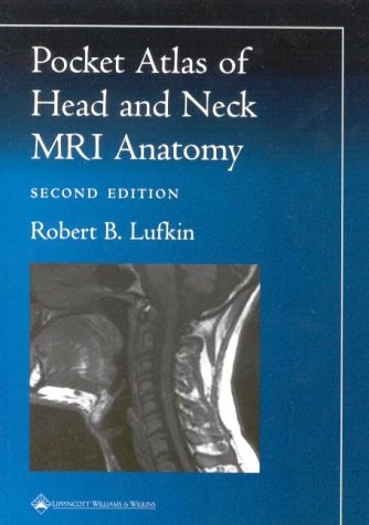
Pocket Atlas of Head and Neck MRI Anatomy
Seiten
2000
|
2nd Revised edition
Lippincott Williams and Wilkins (Verlag)
978-0-7817-2880-5 (ISBN)
Lippincott Williams and Wilkins (Verlag)
978-0-7817-2880-5 (ISBN)
- Titel ist leider vergriffen;
keine Neuauflage - Artikel merken
A guide to head and neck anatomy as seen in state-of-the-art magnetic resonance images. This edition presents high-resolution images of all major areas - the neck, larynx, oropharynx, tongue, nasopharynx, skull base, sinuses, temporal bone, orbit, and temporomandibular joint - displayed in axial, sagittal, and coronal planes.
The thoroughly revised Second Edition of this popular and widely used pocket atlas is a quick, handy guide to head and neck anatomy as seen in state-of-the-art magnetic resonance images. This edition presents 158 new high-resolution images of all major areas - the neck, larynx, oropharynx, tongue, nasopharynx, skull base, sinuses, temporal bone, orbit, and temporomandibular joint - displayed in axial, sagittal, and coronal planes. Anatomic landmarks on each scan are labeled with numbers that correlate to a key at the top of the page. An illustration alongside the key indicates the plane.Praise for the previous edition: "A nice introduction for practicing radiologists who are new to MR of the no-man's land between the skull base and thoracic inlet. Imaging of the head and neck is a growing segment of many radiology practices, and familiarity with this type of normal anatomy is necessary...This is a nice and inexpensive guide to keep at hand in film-viewing areas." - "American Journal of Roentgenology".
The thoroughly revised Second Edition of this popular and widely used pocket atlas is a quick, handy guide to head and neck anatomy as seen in state-of-the-art magnetic resonance images. This edition presents 158 new high-resolution images of all major areas - the neck, larynx, oropharynx, tongue, nasopharynx, skull base, sinuses, temporal bone, orbit, and temporomandibular joint - displayed in axial, sagittal, and coronal planes. Anatomic landmarks on each scan are labeled with numbers that correlate to a key at the top of the page. An illustration alongside the key indicates the plane.Praise for the previous edition: "A nice introduction for practicing radiologists who are new to MR of the no-man's land between the skull base and thoracic inlet. Imaging of the head and neck is a growing segment of many radiology practices, and familiarity with this type of normal anatomy is necessary...This is a nice and inexpensive guide to keep at hand in film-viewing areas." - "American Journal of Roentgenology".
| Erscheint lt. Verlag | 1.9.2000 |
|---|---|
| Reihe/Serie | Radiology Pocket Atlas Series |
| Zusatzinfo | 158 |
| Verlagsort | Philadelphia |
| Sprache | englisch |
| Maße | 124 x 171 mm |
| Gewicht | 118 g |
| Themenwelt | Medizinische Fachgebiete ► Chirurgie ► Neurochirurgie |
| Medizin / Pharmazie ► Medizinische Fachgebiete ► HNO-Heilkunde | |
| Medizinische Fachgebiete ► Radiologie / Bildgebende Verfahren ► Radiologie | |
| ISBN-10 | 0-7817-2880-0 / 0781728800 |
| ISBN-13 | 978-0-7817-2880-5 / 9780781728805 |
| Zustand | Neuware |
| Haben Sie eine Frage zum Produkt? |
Mehr entdecken
aus dem Bereich
aus dem Bereich
Buch | Hardcover (2024)
De Gruyter (Verlag)
CHF 153,90
850 Fakten für die Zusatzbezeichnung
Buch | Softcover (2022)
Springer (Verlag)
CHF 65,80
Buch | Hardcover (2023)
Springer (Verlag)
CHF 307,95


