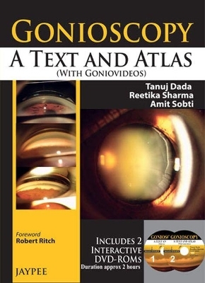
Gonioscopy: A Text and Atlas (with Goniovideos)
Jaypee Brothers Medical Publishers
978-93-5090-434-3 (ISBN)
- Titel ist leider vergriffen;
keine Neuauflage - Artikel merken
Gonioscopy is an eye examination to look at the front part of the eye (anterior chamber), between the cornea and the iris. It is a painless procedure to see whether the area where fluid drains out of the eye is open or closed. Gonioscopy is important in diagnosing and monitoring glaucoma, an eye disease that damages the optic nerve and may result in blindness.
This text and atlas is a step by step guide to train ophthalmologists in the principles, indications, and techniques for performing gonioscopy, whilst highlighting various goniopathologies encountered in different congenital and acquired conditions.
The text begins with the history, principles and indications of gonioscopy, with the following chapters providing an overview of different techniques.
The atlas features more than 400 goniophotographs illustrating different conditions discussed in the text. Also included, are two DVDs that include 40 goniovideos demonstrating various techniques and procedures.
Key points
Step by step guide to gonioscopy
Covers basic principles and indications, and techniques
Features more than 400 goniophotographs
Includes two DVDs demonstrating gonioscopy procedures
Reetika Sharma MD Senior Registrar Amit Sobti MD Senior Registrar All at Dr Rajendra Prasad Centre for Ophthalmic Sciences, All India Institute of Medical Sciences, New Delhi, India
Gonioscopy Text
Chapter 1: History
Chapter 2: Principle of Gonioscopy
Chapter 3: Indications of Gonioscopy
Chapter 4: Direct and Indirect Gonioscopy
Chapter 5: Slit Lamp Gonioscopy Techniques
Chapter 6: Special Gonioscopy Techniques
Chapter 7: Normal Gonioscopy Anatomy
Chapter 8: Gonioscopic Grading Systems
Chapter 9: Assessment of Angle Pathology – An Overview
Chapter 10: Primary Angle Closure Disease
Chapter 11: Primary Open Angle Glaucoma
Chapter 12: Congenital and Developmental Glaucoma
Chapter 13: Angle Pathologies
Chapter 14: Sterilization Techniques
Chapter 15: RetCam Gonioscopy
Gonioscopy Atlas
Open Angle
Myopia
Angle Closure
Closed Angle Opening on Manipulation
Anterior Iris Insertion
Angle Recession
Aniridia
Anterior Chamber Intraocular Lens
Axenfeld-Rieger Syndrome
Blood in the Angle
Ciliary Body Melanoma
Corneal Wedge
Cotton Fibers in the Angle
Cyclodialysis
Express Implant
Fibrous Ingrowth
Foreign Body in the Angle
Goniosynechiae
Haptic of PC IOL in Angle through Iridectomy
Iridodialysis
Irido-fundal Coloboma
Iris Adherent to Cataract Surgical Wound
Iris Bombe
Iris PE Cyst Causing Segmental Angle Closure
Kayser–Fleischer Ring
Neovascularization of Angle
Ocular Ischemic Syndrome
Pigment Dispersion Syndrome
Plateau Iris Configuration with Double Hump Pattern
Post Cataract Surgery Gonioscopy
Post Surgical Angle Pigmentation
Post-traumatic Iridodialysis with Central PAS
Post-traumatic Angle Pigmentation
Prominent Iris Processes
Prominent Schwalbe’s line
Pseudophakia with Glaucoma
Pseudophakia with Glaucoma with AC IOL
Pseudotrabecular Meshwork in a Closed Angle
Sarcoidosis Nodules in the Angle
Secondary Angle Closure due to High Vaulted ICL
Sentinel Synechiae
Silicone Oil
Trabeculectomy Ostium
Uveitic Glaucoma
Vitreous Strands in the Angle
View of Ciliary Body and Peripheral Retina
Ahmed Glaucoma Valve
DVD Contents
Angle Closure Glaucoma
Ahmed Glaucoma Valve
Angle Pigmentation in Pseudophakia
Angle Recession
Aniridia
Anterior Segment Dysgenesis
Axenfled-Rieger Syndrome
Anterior Chamber Iol Causing Angle Damage
Cyclodialysis
Broad Based Peripheral Anterior Synechiae
Foreign Body in Angle
Express Shunt
Fibrous Ingrowth in the Angle
Heavily Pigmented Trabecular Meshwork
Gonioscopy Techniques
High Myope with Wide Open Angle
Irido-Fundal Coloboma
Iridocorneal Endothelial Syndrome
Iridodialysis
Keratic Precipitates
Nodules in the Angle in Sarcoidosis Uveitis
Ocular Squamous Neoplasia Eroding the Angle
Ocular Ischemic Syndrome
Open Angle
Peripheral Retina in Aphakia
Pigment Epithelial Cyst Causing Segmental Angle Closure
Paracytic Cyst
PCIOL in Anterior Chamber Angle
Trabeculectomy Ostium Blockage
Pigment Clumps
Pigment Dispersion
Pigmentary Glaucoma
Tumor in the Angle
Prominent Iris Processes
Silicon Oil in the Angle
Trabeculectomy
Uveitis
Corneal Wedge
Koeppe Lens
Dynamic Gonioscopy
| Erscheint lt. Verlag | 30.6.2013 |
|---|---|
| Zusatzinfo | 400 Halftones, unspecified; 20 Illustrations, unspecified |
| Verlagsort | New Delhi |
| Sprache | englisch |
| Maße | 184 x 241 mm |
| Gewicht | 375 g |
| Themenwelt | Medizin / Pharmazie ► Medizinische Fachgebiete ► Augenheilkunde |
| Studium ► 2. Studienabschnitt (Klinik) ► Anamnese / Körperliche Untersuchung | |
| ISBN-10 | 93-5090-434-9 / 9350904349 |
| ISBN-13 | 978-93-5090-434-3 / 9789350904343 |
| Zustand | Neuware |
| Haben Sie eine Frage zum Produkt? |
aus dem Bereich


