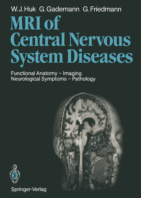
Magnetic Resonance Imaging of Central Nervous System Diseases
Springer Berlin (Verlag)
978-3-642-72570-8 (ISBN)
1 Physical Principles and Techniques of MR Imaging.- 1.1 Physical Principles.- 1.2 Techniques of MR Imaging.- 1.3 Pulse Sequences and Image Contrasts.- 1.4 New Approaches in Clinical MR.- References.- 2 Normal Anatomy of the Central Nervous System.- 2.1 Orientation.- 2.2 Cerebral Cortex.- 2.3 Diencephalon and Basal Ganglia.- 2.4 Midbrain.- 2.5 Posterior Fossa.- 2.6 Optical System.- 2.7 Acoustic System.- 2.8 Cerebral Blood Supply.- 2.9 Spinal Cord.- References.- 3 Practical Aspects of the MR Examination.- 3.1 Preparations.- 3.2 Examination Procedure.- 3.3 Sedation, Anesthesia, and Anesthesiology Monitoring During MR Examinations.- 3.4 Side Effects and Contraindications.- References.- 4 Symptoms and Pathologic Anatomy: A Tabular Listing.- 5 General Aspects of the MR Signal Pattern of Certain Normal and Pathologic Structures.- 5.1 Brain Edema.- 5.2 Intracranial Hemorrhage and Iron Metabolism.- 5.3 Practical Aspects of Blood and CSF Flow.- 5.4 Effects of Radiotherapy on Brain and Spinal Cord Tumors.- 5.5 Effect of Contrast Agent (Gadolinium-DTPA) on the MR Appearance of Normal Tissue.- References.- 6 Magnetic Resonance Imaging of the Brain in Childhood: Development and Pathology.- 6.1 Introduction.- 6.2 Technical Aspects.- 6.3 Normal Appearance.- 6.4 Intracranial Hemorrhage.- 6.5 Infarction.- 6.6 Cysts and Leukomalacias.- 6.7 Hydrocephalus.- 6.8 Congenital Malformations.- 6.9 White Matter Disease.- 6.10 Infection.- 6.11 Delays or Deficits in Myelination.- 6.12 Tumors.- 6.13 Other Diseases.- 6.14 Follow-Up Examination.- 6.15 Conclusion.- References.- 7 Malformations of the CNS.- 7.1 Review of Ontogeny.- 7.2 MR Imaging of CNS Malformations.- 7.3 Midline Closure Defects (Neural Tube Defects, Dysraphic Disorders).- 7.4 Malformations of the Commissures and Midline Structures.- 7.5 Anomalies of Cell Migration.- 7.6 Destructive Anomalies.- 7.7 Neuroectodermal Dysplasias.- 7.8 Miscellaneous Abnormalities.- References.- 8 Degenerative Disorders of the Brain and White Matter Diseases.- 8.1 Primary Neuronal (Gray Matter) Involvement.- 8.2 Primary Myelin (White Matter) Involvement.- 8.3 Primary Involvement of the Basal Ganglia and Brainstem.- 8.4 Primary Involvement with No Sites of Predilection.- References.- 9 Temporal Lobe Epilepsy.- 9.1 MR Findings.- References.- 10 Intracranial Tumors.- 10.1 General Aspects of the Detection of Intracranial Tumors with MR.- 10.2 Tumors of Neuroepithelial Tissue.- 10.3 Tumors of the Nerve Sheath Cells.- 10.4 Tumors of Meningeal and Related Tissues.- 10.5 Primary Malignant Lymphomas.- 10.6 Tumors of Blood Vessel Origin.- 10.7 Germ Cell Tumors.- 10.8 Other Malformative Tumors and Tumor-Like Lesions.- 10.9 Tumors of the Pituitary.- 10.10 Local Extensions from Regional Tumors.- 10.11 Metastatic Tumors.- 10.12 Pseudotumor Cerebri.- References.- 11 Tumors of the Posterior Fossa.- 11.1 Intra-axial Tumors.- 11.2 Extra-axial Tumors.- References.- 12 Diseases of the Eyeball, Orbit, and Accessory Structures.- 12.1 Technical Requirements.- 12.2 Examination Technique.- 12.3 Eyeball.- 12.4 Optic Nerve.- 12.5 Lacrimal Glands.- 12.6 Ocular Muscles.- 12.7 Orbit.- 12.8 Malformations.- 12.9 Trauma.- References.- 13 Vascular Diseases of the Brain.- 13.1 Disturbances of Arterial Blood Flow.- 13.2 Disturbances of Venous Blood Flow.- 13.3 Hemorrhages.- References.- 14 Vascular Malformations.- 14.1 Vascular Malformations of the Brain.- 14.2 Vascular Malformations Involving the Vertebral Canal.- References.- 15 Inflammatory Diseases of the Central Nervous System.- 15.1 General Aspects of the Inflammatory Process.- 15.2 Classification.- 15.3 Infections of the Meninges.- 15.4 Infections of the Brain Tissue.- 15.5 Acquired Immune Deficiency Syndrome.- 15.6 Summary.- References.- 16 Head Injury.- 16.1 Intracranial Extracerebral Hemorrhages.- 16.2 Blunt Brain Injuries.- 16.3 Open Head Injuries.- 16.4 Other Sequelae of Injuries.- 16.5 Late Sequelae of Head Injuries.- References.- 17 Diseases of the Vertebral Column and Spinal Cord.- 17.1 Examination Technique.- 17.2 Intraspinal Processes.- 17.3 Spinal Tumors.- 17.4 Degenerative Diseases of the Spine.- 17.5 Trauma.- References.- Glossary of Magnetic Resonance Terms.
| Erscheint lt. Verlag | 16.12.2011 |
|---|---|
| Mitarbeit |
Assistent: I. Baer, D. Baleriaux, G.M. Bydder, W.L. Curati, M. Deimling, H. Goetz, R. Haerten, W. Heindel, R.H. Hewlett, E. Kraus, J.W. Lotz, G. Michelozzi, U. Mödder, T. Pasch, J.M. Pennock, W. Schajor, W. Steinbrich, H.J. Weinmann, F.E. Zanella |
| Übersetzer | Terry Telger |
| Zusatzinfo | XVI, 452 p. |
| Verlagsort | Berlin |
| Sprache | englisch |
| Maße | 193 x 270 mm |
| Gewicht | 1013 g |
| Themenwelt | Medizinische Fachgebiete ► Radiologie / Bildgebende Verfahren ► Radiologie |
| Schlagworte | Brainstem • classification • Magnetic Resonance Imaging (MRI) • nervous system • radiotherapy • Tumor |
| ISBN-10 | 3-642-72570-8 / 3642725708 |
| ISBN-13 | 978-3-642-72570-8 / 9783642725708 |
| Zustand | Neuware |
| Informationen gemäß Produktsicherheitsverordnung (GPSR) | |
| Haben Sie eine Frage zum Produkt? |
aus dem Bereich


