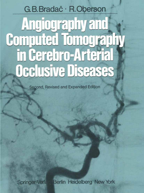
Angiography and Computed Tomography in Cerebro-Arterial Occlusive Diseases
Springer Berlin (Verlag)
978-3-642-68556-9 (ISBN)
1 Etiopathology.- 1.1 Atherosclerosis.- 1.2 Lesions not Due to Atherosclerosis.- 2 Angiography.- 2.1 Indications.- 2.2 Hazards.- 2.3 Technique.- 3 Angiographic Findings.- 3.1 Normal Arteriocerebral Angiograms.- 3.2 Lesions of the Extracranial Segments of the Cerebral Arteries.- 3.3 Lesions of the Carotid Siphon.- 3.4 Lesions in the Region of the Middle Cerebral Artery.- 3.5 Lesions of the Posterior Cerebral and Basilar Arteries.- 3.6 Other Pathologic Findings in the Vertebrobasilar System.- 3.7 Lesions in the Region of the Anterior Cerebral Artery.- 3.8 Lesions in the Region of the Anterior Choroidal Artery.- 3.9 Lesions in the Region of the Lenticulostriate Arteries.- 3.10 Rare Lesions of the Intracranial Vessels.- 3.11 Collateral Flow.- 3.12 The Negative Angiogram.- 3.13 Indication and Modalities of Surgical Therapy.- 4 Computed Tomography in the Diagnosis of Cerebrovascular Occlusive Diseases.- 4.1 Patients with Transient Ischemic Attacks.- 4.2 Patients with Completed Stroke.- 5 Other Investigations in the Diagnosis of Cerebrovascular Occlusive Diseases.- 5.1 Carotid Auscultation.- 5.2 Ophthalmodynamometry.- 5.3 Doppler Ultrasound.- 5.4 Radionuclide Brain Scan.- 5.5 Regional Central Blood Flow Measurements.- 5.6 Intravenous Angiography.- 6 Some Conclusive Considerations on the Pathogenesis of TIAs and Infarctions.- 6.1 TIA in the Carotid Sector.- 6.2 TIA in the Vertebrobasilar Sector.- 6.3 Infarction.- 7 Conclusions on the Use of Diagnostic Procedures.- References.
| Erscheint lt. Verlag | 21.11.2011 |
|---|---|
| Vorwort | J.-M. Taveras |
| Zusatzinfo | XII, 290 p. |
| Verlagsort | Berlin |
| Sprache | englisch |
| Maße | 210 x 280 mm |
| Gewicht | 756 g |
| Themenwelt | Medizinische Fachgebiete ► Chirurgie ► Neurochirurgie |
| Medizinische Fachgebiete ► Innere Medizin ► Kardiologie / Angiologie | |
| Medizinische Fachgebiete ► Radiologie / Bildgebende Verfahren ► Radiologie | |
| Schlagworte | Angiography • Arteries • brain • Circulation • Computed tomography (CT) • embolism • Tomography • vascular disease |
| ISBN-10 | 3-642-68556-0 / 3642685560 |
| ISBN-13 | 978-3-642-68556-9 / 9783642685569 |
| Zustand | Neuware |
| Informationen gemäß Produktsicherheitsverordnung (GPSR) | |
| Haben Sie eine Frage zum Produkt? |
aus dem Bereich


