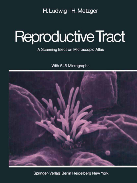
The Human Female Reproductive Tract
Springer Berlin (Verlag)
978-3-642-66347-5 (ISBN)
Materials.- Methodology.- 1. The Vagina.- Tissue Surface of the Vaginal Epithelium.- 2. The Ectocervix and Endocervix.- Transition Zone between the Ectocervix and the Endocervix.- Ectocervix.- Endocervix.- 3. The Endometrium.- Gross Architecture of the Endometrial Gland Openings.- Cellular Shape of the Endometrial Surface Epithelium around the Gland Openings.- Endometrial Surface Epithelium.- Cellular Details of the Endometrial Surface Epithelium.- Comparative Micromorphology of Ciliated Cells in the Endometrium.- Comparative Micromorphology of the Microvillous Relief of the Nonciliated Cells in the Endometrium.- The Re-Epithelization of the Postmenstrual Uterine Cavity.- Endometrium after the Insertion of IUD.- Endometrium under the Influence of Ethinylestradiol.- Endometrium of a Female Fetus (Week 23 of Pregnancy).- Senile Endometrium.- 4. The Fallopian Tube.- Organization of the Ampullary Mucosa of the Oviduct.- Gross Arrangement of Ciliated and Nonciliated Cells in the Ampulla.- Distribution of Ciliated and Nonciliated Cells in the Ampulla.- Relation between Single Ciliated and Nonciliated Cells in the Ampulla.- Boundaries of the Nonciliated Cells Occurring in the Ampulla.- Microvilli of Nonciliated Cells Occurring in the Ampulla.- Comparative Topology of the Segments in the Oviduct.- Fimbriae of a Female Fetus (Week 23 of Pregnancy).- 5. The Ovary.- The Surface of an Adult Ovary at the Time of Ovulation.- The Surface of an Adult Ovary during the Luteal Phase.- The Texture of the Second Layer of the Tunica Albuginea.- 6. Gestational Metamorphosis of the Tissue Surface.- Vagina (Pregnancy).- Ectocervix (Pregnancy).- Endocervix (Pregnancy).- Lower Uterine Segment (Pregnancy).- Endometrium (Pregnancy).- The Oviduct (Ectopic Pregnancy).- The Oviduct (at Term of Pregnancy).- 7. Metamorphosis of the Tissue Surface by Progestational Agents.- Ectocervix (Progestogenic Treatment).- Endocervix (Progestogenic Treatment).- Endometrium (Progestogenic Treatment).- The Oviduct (Progestogenic Treatment).- 8. The Placenta.- Organization of the Villous Tree.- Branching of the Placental Villous Tree.- Microvillous Pattern of the Terminal Villi.- Details of the Microvillous Pattern of Terminal Villi.- Microvillous Pattern around a Placental Sprout.- The Basal Plate.- Surface of the Syncytiotrophoblast in Toxemia.- Infarction of the Intervillous Space in Toxemia.- 9. The Membranes.- Organization of the Fetal Surface of the Amniotic Epithelium.- Arrangement of the Amniotic Epithelial Cells.- Surface Pattern and Cellular Shape of Amniotic Epithelial Cells.- Surface Details of the Amniotic Epithelium.- Microvilli of the Amniotic Epithelium.- The Fibroelastic Layer of the Amnion (Amnion Seen from the Chorionic Side after Removal of Chorion).- Surface of Amniotic Epithelium in Blood Group Incompatibility.- Surface of Amniotic Epithelium in Postmaturity.- Conclusions.- References.
| Erscheint lt. Verlag | 15.11.2011 |
|---|---|
| Zusatzinfo | XIV, 250 p. |
| Verlagsort | Berlin |
| Sprache | englisch |
| Maße | 210 x 280 mm |
| Gewicht | 662 g |
| Themenwelt | Studium ► 1. Studienabschnitt (Vorklinik) ► Anatomie / Neuroanatomie |
| Schlagworte | Elektronenmikroskopie • growth • Obstetrics • Reproductive Tract • Weibliches Geschlechtsorgan |
| ISBN-10 | 3-642-66347-8 / 3642663478 |
| ISBN-13 | 978-3-642-66347-5 / 9783642663475 |
| Zustand | Neuware |
| Haben Sie eine Frage zum Produkt? |
aus dem Bereich


