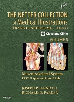
Netter Collection of Medical Illustrations: Musculoskeletal System
Saunders (Verlag)
978-1-4160-6382-7 (ISBN)
- Titel ist leider vergriffen;
keine Neuauflage - Artikel merken
"The Lower Limb and Spine, Part 2" of "The Netter Collection of Medical Illustrations: Musculoskeletal System, 2nd Edition", provides a highly visual guide to the spine and lower extremity, from basic science and anatomy to orthopaedics and rheumatology. This spectacularly illustrated volume in the masterwork known as the (CIBA) "Green Books" has been expanded and revised by Dr. Joseph Iannotti, Dr. Richard Parker, and other experts from the Cleveland Clinic to mirror the many exciting advances in musculoskeletal medicine and imaging - offering rich insights into the anatomy, physiology, and clinical conditions of the spine; pelvis, hip, and thigh; knee; lower leg; and ankle and foot.
SECTION 1-SPINE 1-1 Vertebral Column, 2 Cervical Spine 1-2 Atlas and Axis, 3 1-3 External Craniocervical Ligaments, 4 1-4 Internal Craniocervical Ligaments, 5 1-5 Suboccipital Triangle, 6 1-6 Dens Fracture, 7 1-7 Jefferson and Hangman's Fractures, 8 1-8 Cervical Vertebrae, 9 1-9 Muscles of Back: Superficial Layers, 10 1-10 Muscles of Back: Intermediate and Deep Layers, 11 1-11 Spinal Nerves and Sensory Dermatomes, 12 1-12 Cervical Spondylosis, 13 1-13 Cervical Spondylosis and Myelopathy, 14 1-14 Cervical Disc Herniation: Clinical Manifestations, 15 1-15 Surgical Approaches for the Treatment of Myelopathy and Radiculopathy, 16 1-16 Extravascular Compression of Vertebral Arteries, 17 Thoracolumbar and Sacral Spine 1-17 Thoracic Vertebrae and Ligaments, 18 1-18 Lumbar Vertebrae and Intervertebral Discs, 19 1-19 Sacral Spine and Pelvis, 20 1-20 Lumbosacral Ligaments, 21 1-21 Degenerative Disc Disease, 22 1-22 Lumbar Disc Herniation, 23 1-23 Lumbar Spinal Stenosis, 24 1-24 Lumbar Spinal Stenosis (Continued), 25 1-25 Degenerative Lumbar Spondylolisthesis, 26 1-26 Degenerative Spondylolisthesis: Cascading Spine, 27 1-27 Adult Deformity, 28 1-28 Three-Column Concept of Spinal Stability and Compression Fractures, 29 1-29 Compression Fractures (Continued), 30 1-30 Burst, Chance, and Unstable Fractures, 31 Deformities of Spine 1-31 Congenital Anomalies of Occipitocervical Junction, 32 1-32 Congenital Anomalies of Occipitocervical Junction (Continued), 33 1-33 Synostosis of Cervical Spine (Klippel-Feil Syndrome), 34 1-34 Clinical Appearance of Congenital Muscular Torticollis (Wryneck), 35 1-35 Nonmuscular Causes of Torticollis, 36 1-36 Pathologic Anatomy of Scoliosis, 37 1-37 Typical Scoliosis Curve Patterns, 38 1-38 Congenital Scoliosis: Closed Vertebral Types (MacEwen Classification), 39 1-39 Clinical Evaluation of Scoliosis, 40 1-40 Determination of Skeletal Maturation, Measurement of Curvature, and Measurement of Rotation, 41 1-41 Braces for Scoliosis, 42 1-42 Scheuermann Disease, 43 1-43 Congenital Kyphosis, 44 1-44 Spondylolysis and Spondylolisthesis, 45 1-45 Myelodysplasia, 46 1-46 Lumbosacral Agenesis, 47 SECTION 2-PELVIS, HIP, AND THIGH Anatomy 2-1 Superficial Veins and Cutaneous Nerves, 50 2-2 Lumbosacral Plexus, 52 2-3 Sacral and Coccygeal Plexuses, 53 2-4 Nerves of Buttock, 54 2-5 Femoral Nerve (L2, 3, 4) and Lateral Femoral Cutaneous Nerve (L2, 3), 55 2-6 Obturator Nerve (L2, 3, 4), 56 2-7 Sciatic Nerve (L4, 5; S1, 2, 3) and Posterior Femoral Cutaneous Nerve (S1, 2, 3), 57 2-8 Muscles of Front of Hip and Thigh, 58 2-9 Muscles of Hip and Thigh (Anterior and Lateral Views), 59 2-10 Muscles of Back of Hip and Thigh, 60 2-11 Bony Attachments of Muscles of Hip and Thigh: Anterior View, 61 2-12 Bony Attachments of Muscles of Hip and Thigh: Posterior View, 62 2-13 Cross-Sectional Anatomy of Hip: Axial View, 63 2-14 Cross-Sectional Anatomy of Hip: Coronal View, 64 2-15 Cross-Sectional Anatomy of Thigh, 65 2-16 Arteries and Nerves of Thigh: Anterior Views, 66 2-17 Arteries and Nerves of Thigh: Deep Dissection (Anterior View), 67 2-18 Arteries and Nerves of Thigh: Deep Dissection (Posterior view), 68 2-19 Bones and Ligaments at Hip: Osteology of the Femur, 69 2-20 Bones and Ligaments at Hip: Hip Joint, 70 Physical Examination 2-21 Physical Examination, 71 Deformities of the Pelvis and Femur 2-22 Proximal Femoral Focal Deficiency: Radiographic Classification, 72 2-23 Proximal Femoral Focal Deficiency: Clinical Presentation, 73 2-24 Congenital Short Femur with Coxa Vara, 74 2-25 Recognition of Developmental Dislocation of the Hip, 75 2-26 Clinical Findings in Developmental Dislocation of Hip, 76 2-27 Radiologic Diagnosis of Developmental Dislocation of Hip, 77 2-28 Adaptive Changes in Dislocated Hip That Interfere with Reduction, 78 2-29 Device for Treatment of Clinically Reducible Dislocation of Hip, 79 2-30 Blood Supply to Femoral Head in Infancy, 80 2-31 Legg-Calve-Perthes Disease: Pathogenesis, 81 2-32 Legg-Calve-Perthes Disease: Physical Examination, 82 2-33 Legg-Calve-Perthes Disease: Physical Examination (Continued), 83 2-34 Stages of Legg-Calve-Perthes Disease, 84 2-35 Legg-Calve-Perthes Disease: Lateral Pillar Classification, 85 2-36 Legg-Calve-Perthes Disease: Conservative Management, 86 2-37 Femoral Varus Derotational Osteotomy, 87 2-38 Innominate Osteotomy, 88 2-39 Innominate Osteotomy (Continued), 89 2-40 Physical Examination and Classification of Slipped Capital Femoral Epiphysis, 90 2-41 Pin Fixation in Slipped Capital Femoral Epiphysis, 91 Disorders of the Hip 2-42 Hip Joint Involvement in Osteoarthritis, 92 2-43 Total Hip Replacement: Prostheses, 93 2-44 Total Hip Replacement: Steps 1 to 3, 94 2-45 Total Hip Replacement: Steps 4 to 8, 95 2-46 Total Hip Replacement: Steps 9 to 12, 96 2-47 Total Hip Replacement: Steps 13 to 18, 97 2-48 Total Hip Replacement: Steps 19 and 20, 98 2-49 Total Hip Replacement: Dysplastic Acetabulum, 99 2-50 Total Hip Replacement: Protrusio Acetabuli, 100 2-51 Total Hip Replacement: Complications- Loosening of Femoral Component, 101 2-52 Total Hip Replacement: Complications- Fractures of Femur and Femoral Component, 102 2-53 Total Hip Replacement: Complications- Loosening of Acetabular Component and Dislocation of Total Hip Prosthesis, 103 2-54 Total Hip Replacement: Infection, 104 2-55 Total Hip Replacement: Hemiarthroplasty of Hip, 105 2-56 Hip Resurfacing, 106 2-57 Rehabilitation after Total Hip Replacement, 107 2-58 Femoroacetabular Impingement/ Hip Labral Tears, 108 2-59 Avascular Necrosis, 109 2-60 Trochanteric Bursitis, 110 2-61 Snapping Hip (Coxa Saltans), 111 2-62 Muscle Strains, 112 Trauma 2-63 Injury to Pelvis: Stable Pelvic Ring Fractures, 113 2-64 Injury to Pelvis: Straddle Fracture and Lateral Compression Injury, 114 2-65 Injury to Pelvis: Open Book Fracture, 115 2-66 Injury to Pelvis: Vertical Shear Fracture, 116 2-67 Injury to Hip: Acetabular Fractures, 117 2-68 Injury to Hip: Acetabular Fractures (Continued), 118 2-69 Injury to Hip: Posterior Dislocation of Hip, 119 2-70 Injury to Hip: Anterior Dislocation of Hip, Obturator Type, 120 2-71 Injury to Hip: Dislocation of Hip with Fracture of Femoral Head, 121 2-72 Injury to Femur: Intracapsular Fracture of Femoral Neck, 122 2-73 Injury to Femur: Intertrochanteric Fracture of Femur, 123 2-74 Injury to Femur: Subtrochanteric Fracture of Femur, 124 2-75 Injury to Femur: Fracture of Shaft of Femur, 125 2-76 Injury to Femur: Fracture of Distal Femur, 126 2-77 Amputation of Lower Limb and Hip (Disarticulation and Hemipelvectomy), 127 SECTION 3-KNEE Anatomy 3-1 Topographic Anatomy of the Knee, 130 3-2 Osteology of the Knee, 131 3-3 Knee: Lateral and Medial Views, 132 3-4 Knee: Anterior Views, 133 3-5 Knee: Posterior and Sagittal Views, 134 3-6 Knee: Interior View and Cruciate and Collateral Ligaments, 135 3-7 Arteries and Nerves of Knee, 136 Injury to the Knee 3-8 Arthrocentesis of Knee Joint, 137 3-9 Types of Meniscal Tears and Discoid Meniscus Variations, 138 3-10 Tears of the Meniscus, 139 3-11 Medial and Lateral Meniscus, 140 3-12 Rupture of the Anterior Cruciate Ligament, 141 3-13 Lateral Pivot Shift Test for Anterolateral Knee Instability, 142 3-14 Rupture of Cruciate Ligaments: Arthroscopy, 143 3-15 Rupture of Posterior Cruciate Ligament, 144 3-16 Physical Examination of the Leg and Knee, 145 3-17 Sprains of Knee Ligaments, 146 3-18 Disruption of Quadriceps Femoris Tendon or Patellar Ligament, 147 3-19 Dislocation of Knee Joint, 148 Disorders of the Knee 3-20 Progression of Osteochondritis Dissecans, 149 3-21 Osteonecrosis, 150 3-22 Tibial Intercondylar Eminence Fracture, 151 3-23 Synovial Plica, 152 3-24 Synovial Plica (Arthroscopy), Bursitis, and Iliotibial Band Friction Syndrome, 153 3-25 Pigmented Villonodular Synovitis and Meniscal Cysts, 154 3-26 Rehabilitation after Injury to Knee Ligaments, 155 3-27 Bipartite Patella and Baker's Cyst, 156 3-28 Subluxation and Dislocation of Patella, 157 3-29 Fracture of the Patella, 158 3-30 Osgood-Schlatter Lesion, 159 3-31 Knee Arthroplasty: Osteoarthritis of the Knee, 160 3-32 Knee Arthroplasty: Total Condylar Prosthesis and Unicompartmental Prosthesis, 161 3-33 Knee Arthroplasty: Posterior Stabilized Knee Prosthesis, 162 3-34 Total Knee Replacement Technique: Steps 1 to 5, 163 3-35 Total Knee Replacement Technique: Steps 6 to 9, 164 3-36 Total Knee Replacement Technique: Steps 10 to 14, 165 3-37 Total Knee Replacement Technique: Steps 15 to 20, 166 3-38 Medial Release for Varus Deformity of Knee, 167 3-39 Lateral Release for Valgus Deformity of Knee, 168 3-40 Rehabilitation after Total Knee Replacement, 169 3-41 High Tibial Osteotomy for Varus Deformity of Knee, 170 3-42 Below-Knee Amputation, 171 3-43 Disarticulation of Knee and Above-Knee Amputation, 172 SECTION 4-LOWER LEG Anatomy 4-1 Topographic Anatomy of the Lower Leg, 174 4-2 Fascial Compartments of Leg, 175 4-3 Muscles of Leg: Superficial Dissection (Anterior View), 176 4-4 Muscles of Leg: Superficial Dissection (Lateral View), 177 4-5 Muscles, Arteries, and Nerves of Leg: Deep Dissection (Anterior View), 178 4-6 Muscles of Leg: Superficial Dissection (Posterior View), 179 4-7 Muscles of Leg: Intermediate Dissection (Posterior View), 180 4-8 Muscles, Arteries, and Nerves of Leg: Deep Dissection (Posterior View), 181 4-9 Common Peroneal Nerve, 182 4-10 Tibial Nerve, 183 4-11 Tibia and Fibula, 184 4-12 Tibia and Fibula (Continued), 185 4-13 Bony Attachments of Muscles of Leg, 186 Injury to Lower Leg 4-14 Fracture of Proximal Tibia Involving Articular Surface, 187 4-15 Fracture of Shaft of Tibia, 188 4-16 Fracture of Tibia in Children, 189 Congenital Deformities 4-17 Bowleg and Knock-Knee, 190 4-18 Blount Disease, 191 4-19 Toeing In: Metatarsus Adductus and Internal Tibial Torsion, 192 4-20 Toeing In: Internal Femoral Torsion, 193 4-21 Toeing Out and Postural Torsional Effects on Lower Limbs, 194 SECTION 5-ANKLE AND FOOT Anatomy 5-1 Surface Anatomy and Muscle Origins and Insertions, 196 5-2 Tendon Sheaths of Ankle, 197 5-3 Ligaments and Tendons of Ankle, 198 5-4 Dorsal Foot: Superficial Dissection, 199 5-5 Dorsal Foot: Deep Dissection, 200 5-6 Plantar Foot: Superficial Dissection, 201 5-7 Plantar Foot: First Layer, 202 5-8 Plantar Foot: Second Layer, 203 5-9 Plantar Foot: Third Layer, 204 5-10 Interosseous Muscles and Deep Arteries of Foot, 205 5-11 Cross-Sectional Anatomy of Ankle and Foot, 206 5-12 Cross-Sectional Anatomy of Ankle and Foot (Continued), 207 5-13 Bones of Foot, 208 5-14 Bones of Foot (Continued), 209 5-15 Ligaments and Tendons of Foot: Plantar View, 210 5-16 Lymph Vessels and Nodes of Lower Limb, 211 Fractures and Dislocations 5-17 Major Sprains and Sprain Fractures, 212 5-18 Mechanisms of Ankle Sprains, 213 5-19 Rotational Fractures, 214 5-20 Repair of Fracture of Malleolus, 215 5-21 Pilon Fracture, 216 5-22 Talus Fracture, 217 5-23 Extra-articular Fracture of Calcaneus, 218 5-24 Intra-articular Fracture of Calcaneus, 219 5-25 Fifth Metatarsal Fractures, 220 5-26 Lisfranc Injury, 221 5-27 Navicular Stress Fractures, 222 Common Soft Tissue Disorders 5-28 Achilles Tendon Rupture, 223 5-29 Peroneal Tendon Injury, 224 5-30 Osteochondral Lesions of the Talus, 225 5-31 Turf Toe, 226 5-32 Plantar Fasciitis, 227 5-33 Posterior Tibial Tendonitis/Flatfoot, 228 Deformities of the Ankle and Foot 5-34 Congenital Clubfoot, 229 5-35 Congenital Clubfoot (Continued), 230 5-36 Congenital Vertical Talus, 231 5-37 Cavovarus Foot, 232 5-38 Calcaneovalgus and Planovalgus, 233 5-39 Tarsal Coalition, 234 5-40 Tarsal Coalition (Continued), 235 5-41 Accessory Tarsal Navicular, 236 5-42 Congenital Toe Deformities, 237 5-43 Kohler Disease, 238 Infections and Amputations 5-44 Common Foot Infections, 239 5-45 Deep Infections of Foot, 240 5-46 Lesions of the Diabetic Foot, 241 5-47 Clinical Evaluation of Patient with Diabetic Foot Lesion, 242 5-48 Amputation of Foot, 243 5-49 Syme Amputation (Wagner Modification), 244
| Reihe/Serie | Netter Green Book Collection |
|---|---|
| Zusatzinfo | Approx. 300 illustrations (300 in full color) |
| Verlagsort | Philadelphia |
| Sprache | englisch |
| Maße | 246 x 297 mm |
| Gewicht | 1315 g |
| Themenwelt | Medizin / Pharmazie ► Medizinische Fachgebiete ► Orthopädie |
| Studium ► 1. Studienabschnitt (Vorklinik) ► Anatomie / Neuroanatomie | |
| ISBN-10 | 1-4160-6382-X / 141606382X |
| ISBN-13 | 978-1-4160-6382-7 / 9781416063827 |
| Zustand | Neuware |
| Informationen gemäß Produktsicherheitsverordnung (GPSR) | |
| Haben Sie eine Frage zum Produkt? |
aus dem Bereich


