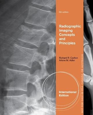
Radiographic Imaging Concepts and Principles, International Edition
Delmar Cengage Learning (Verlag)
978-1-111-31081-3 (ISBN)
- Titel z.Zt. nicht lieferbar
- Versandkostenfrei
- Auch auf Rechnung
- Artikel merken
Arlene Adler served four decades as a Professor in the Radiologic Sciences Programs at Indiana University Northwest. Now Professor Emerita, Ms. Adler authored several articles and textbooks on the radiologic sciences during her career. A member of the American Society of Radiologic Technologists and Fellow of the Association of Educators in Radiologic Sciences (A.E.I.R.S.), she earned her B.S. in Radiologic Sciences from the University of Health Sciences/Chicago Medical School and an M.S. in Education from the University of Illinois. Richard Carlton is an Associate Professor and former Director of Radiologic and Imaging Sciences at Grand Valley State University in Grand Rapids, Michigan. Previously, he taught Radiography at Arkansas State University, City College of San Francisco and Lima Technical College, and worked as a Clinical Coordinator at Wilbur Wright Community College. A Charter Fellow and past president of A.E.I.R.S., Mr. Carlton gives presentations around the world and founded Lambda Nu, the national honor society for radiologic and imaging sciences. He has written 36 books, started two professional journals and served as a J.R.C.E.R.T. site visitor for forty years. Mr. Carlton earned an M.S. from National Louis University, a B.S. from The Chicago Medical School and an A.A.S. from Illinois Central College. He is A.R.R.T. certified in C.V. and radiography and continues to train residents at West Michigan Regional Laboratory in Grand Rapids, Michigan.
1. Mathematics Review.
UNIT I CREATION OF X-RAYS.
2. The Concept of Radiation.
3. Electrical Components.
4. Electromagnetic Components.
5. Radiographic Equipment.
6. X-ray Tubes.
7. Production of X-rays.
UNIT II RADIATION PROTECTION: THE BASICS
8. Radiation Protection Concepts & Equipment.
9. Radiation Protection for Patients and Personnel.
10. Filters.
11. Prime Factors.
12. Interactions between X-rays and Matter.
13. Reducing Patient Dose.
UNIT III THE RADIOGRAPHIC IMAGE.
14. Visual Factors and Perception.
15. Collimation/Beam Restriction.
16. The Patient.
17. Pathology.
18. Grids.
19. Film.
20. Film Wet Chemical Processing.
21. Sensitometry.
22. Film/Screen Combinations.
23. Digital Imaging.
24. PACS Systems.
UNIT IV RADIOGRAPHIC IMAGE ANALYSIS.
25. The Process.
26. Density/Image Receptor Exposure.
27. Contrast.
28. Recorded Detail.
29. Distortion.
30. Image Critique.
31. Quality Assurance/Quality Control.
UNIT V COMPARISON OF EXPOSURE SYSTEMS.
32. Exposure Systems.
33. Automatic Exposure Controls.
34. Exposure Problems.
UNIT VI ADVANCED MODALITIES.
35. Mobile Radiographic Imaging.
36. Fluoroscopic Equipment.
37. Tomographic Equipment & Digital Tomosynthesis.
38. Mammographic Equipment.
39. Bone Densitometry Equipment.
40. Vascular Imaging Instrumentation.
41. Computed Tomography Instrumentation.
42. Magnetic Resonance Imaging Instrumentation.
43. Nuclear Medicine and PET Scanning Instrumentation.
44. Radiation Therapy and Medical Dosimetry Instrumentation.
45. Diagnostic Medical Sonography Instrumentation.
| Verlagsort | Clifton Park |
|---|---|
| Sprache | englisch |
| Maße | 187 x 235 mm |
| Gewicht | 1225 g |
| Themenwelt | Medizin / Pharmazie ► Gesundheitsfachberufe ► MTA - Radiologie |
| Medizin / Pharmazie ► Medizinische Fachgebiete ► Radiologie / Bildgebende Verfahren | |
| ISBN-10 | 1-111-31081-5 / 1111310815 |
| ISBN-13 | 978-1-111-31081-3 / 9781111310813 |
| Zustand | Neuware |
| Haben Sie eine Frage zum Produkt? |
aus dem Bereich


