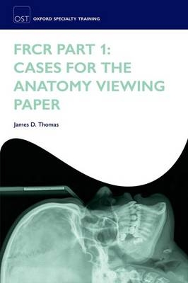
FRCR Part 1: Cases for the anatomy viewing paper
Seiten
2011
Oxford University Press (Verlag)
978-0-19-960453-1 (ISBN)
Oxford University Press (Verlag)
978-0-19-960453-1 (ISBN)
- Titel ist leider vergriffen;
keine Neuauflage - Artikel merken
Exclusively focused on preparing candidates for the FRCR Part 1 anatomy viewing paper, this book enables them to practice questions that have the look and feel of the actual exam. Containing eight practice examinations, the questions are pitched at increasing levels of difficulty.
Exclusively focused on preparing candidates for the FRCR Part 1 anatomy viewing paper, this book enables them to practice questions that have the look and feel of the actual exam. Containing eight practice examinations, each with 20 cases which have been thoroughly reviewed and tested by radiology registrars who have sat the exam, the questions are at increasing levels of difficulty. Screenshots from Osirix and advice on how to approach the exam familiarize candidates with its format. Each exam in the book contains a wide selection of images with all body parts and modalities equally represented to thoroughly test candidates interpretation skills. The 160 images cover all major plain films, CT, MRI, barium studies and other contrast examinations, as well as some of the newer techniques, based on the examples published online by the Royal College of Radiologists.
Exclusively focused on preparing candidates for the FRCR Part 1 anatomy viewing paper, this book enables them to practice questions that have the look and feel of the actual exam. Containing eight practice examinations, each with 20 cases which have been thoroughly reviewed and tested by radiology registrars who have sat the exam, the questions are at increasing levels of difficulty. Screenshots from Osirix and advice on how to approach the exam familiarize candidates with its format. Each exam in the book contains a wide selection of images with all body parts and modalities equally represented to thoroughly test candidates interpretation skills. The 160 images cover all major plain films, CT, MRI, barium studies and other contrast examinations, as well as some of the newer techniques, based on the examples published online by the Royal College of Radiologists.
Dr Thomas is currently a registrar in radiology at Nottingham University Hospitals. He is the co-author of the Oxford Handbook of Clinical Examination and Practical Skills as well as the three sets of Oxford Handbooks Clinical Tutor Study Cards.
Introduction ; EXAM 1 ; Questions ; Answers ; EXAM 2 ; Questions ; Answers ; EXAM 3 ; Questions ; Answers ; EXAM 4 ; Questions ; Answers ; EXAM 5 ; Questions ; Answers ; EXAM 6 ; Questions ; Answers ; EXAM 7 ; Questions ; Answers ; EXAM 8 ; Questions ; Answers
| Erscheint lt. Verlag | 3.11.2011 |
|---|---|
| Verlagsort | Oxford |
| Sprache | englisch |
| Gewicht | 396 g |
| Themenwelt | Medizinische Fachgebiete ► Radiologie / Bildgebende Verfahren ► Radiologie |
| Studium ► 1. Studienabschnitt (Vorklinik) ► Anatomie / Neuroanatomie | |
| ISBN-10 | 0-19-960453-3 / 0199604533 |
| ISBN-13 | 978-0-19-960453-1 / 9780199604531 |
| Zustand | Neuware |
| Haben Sie eine Frage zum Produkt? |
Mehr entdecken
aus dem Bereich
aus dem Bereich
Buch (2023)
Thieme (Verlag)
CHF 265,95


