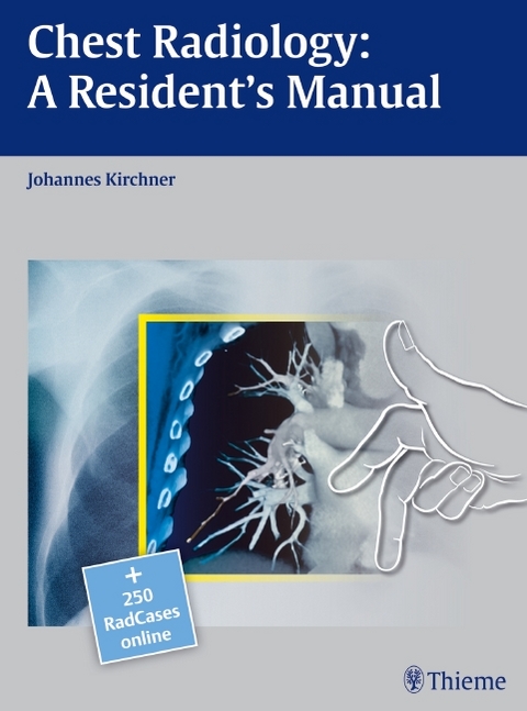
Chest Radiology: A Resident's Manual
Seiten
2011
Thieme (Verlag)
978-3-13-153871-0 (ISBN)
Thieme (Verlag)
978-3-13-153871-0 (ISBN)
lt;br>Learn the essentials of diagnostic chest radiology with this concisely written and visually stunning manual
Chest Radiology: A Resident's Manual is a comprehensive introduction to reading and analyzing radiologic cardiopulmonary images. Readers are guided through systemic image analysis and can further enhance their learning experience with training cases found at the end of each chapter. Cases describe and discuss frequently asked questions regarding heart failure, bronchitis, pneumonia, bronchial carcinoma, fibrosis, pleural disorders, and more. This user-friendly manual will allow the reader to confidently answer the most important and commonly encountered questions related to plain chest radiographs in daily clinical practice. The easy-to-read layout pairs explanatory text on the left page with related drawings and images on the right, allowing readers to navigate their way through each section with ease.
Features
- More than 600 high-resolution images and illustrations demonstrate a wealth of pathology- Concise descriptions explain how to examine conventional x-ray and CT images- Numerous callout boxes in each chapter highlight key takeaway points- A scratch-off code provides access to a searchable online database of 250 must-know thoracic imaging cases
This practice-oriented manual is an invaluable resource and reference guide for residents and radiologists-in-training.
lt;p>1 Heart Failure
2 Bronchitis
3 Pneumonia
4 Bronchial Carcinoma
5 Fibrosing Lung Disease
6 Pleura
| Erscheint lt. Verlag | 9.3.2011 |
|---|---|
| Verlagsort | Stuttgart |
| Sprache | englisch |
| Maße | 230 x 310 mm |
| Gewicht | 1248 g |
| Themenwelt | Medizinische Fachgebiete ► Radiologie / Bildgebende Verfahren ► Radiologie |
| Schlagworte | Acute bronchitis • alveolar cell carcinoma • alveolar pneumonia • Bildgebende Verfahren • bronchial carcinoma • Bronchiectasis • Bronchitis • Brustkorb • Brustorgane • cardiopulmonary image • Chest • chest organs • Chest Radiograph • Chest Radiology • chronic bronchitis • Fibrosis • heart • Heart Failure • Herz • Humanmedizin • Image Analysis • imaging procedures • left heart enlargement • left heart failure • Lung • Lunge • Pleural Disorders • pleural effusion • Pneumonia • Pulmonary Emphysema • Radiologie • Radiologie; Atlanten • Radiologie; Atlas • Radiology • right heart enlargement • right heart failure • thoracic image • Tuberculosis |
| ISBN-10 | 3-13-153871-6 / 3131538716 |
| ISBN-13 | 978-3-13-153871-0 / 9783131538710 |
| Zustand | Neuware |
| Haben Sie eine Frage zum Produkt? |
Mehr entdecken
aus dem Bereich
aus dem Bereich
Buch (2023)
Thieme (Verlag)
CHF 265,95


