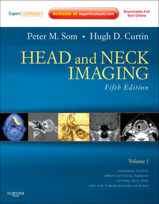
Head and Neck Imaging
Seiten
2011
|
5th Revised edition
Mosby
978-0-323-05355-6 (ISBN)
Mosby
978-0-323-05355-6 (ISBN)
Features imaging examples that help you recognize the imaging presentation of the full range of head and neck disorders using PET, CT, MRI, and ultrasound. This title covers the complexities of embryology, anatomy, and physiology, including anatomical images that help you distinguish subtle abnormalities and understand their etiologies.
"Head and Neck Imaging", by Drs. Peter M. Som and Hugh D. Curtin, delivers the encyclopedic and authoritative guidance you've come to expect from this book - the expert guidance you need to diagnose the most challenging disorders using today's most accurate techniques. New state-of-the-art imaging examples throughout help you recognize the imaging presentation of the full range of head and neck disorders using PET, CT, MRI, and ultrasound. Enhanced coverage of the complexities of embryology, anatomy, and physiology, including original color drawings and new color anatomical images from Frank Netter, help you distinguish subtle abnormalities and understand their etiologies. Access to the complete book's contents is available online at our associated website, which allows you to compare its images onscreen with the imaging findings you encounter in practice.
"Head and Neck Imaging", by Drs. Peter M. Som and Hugh D. Curtin, delivers the encyclopedic and authoritative guidance you've come to expect from this book - the expert guidance you need to diagnose the most challenging disorders using today's most accurate techniques. New state-of-the-art imaging examples throughout help you recognize the imaging presentation of the full range of head and neck disorders using PET, CT, MRI, and ultrasound. Enhanced coverage of the complexities of embryology, anatomy, and physiology, including original color drawings and new color anatomical images from Frank Netter, help you distinguish subtle abnormalities and understand their etiologies. Access to the complete book's contents is available online at our associated website, which allows you to compare its images onscreen with the imaging findings you encounter in practice.
| Zusatzinfo | Approx. 4250 illustrations (250 in full color) |
|---|---|
| Verlagsort | St Louis |
| Sprache | englisch |
| Maße | 222 x 281 mm |
| Gewicht | 9693 g |
| Themenwelt | Medizinische Fachgebiete ► Radiologie / Bildgebende Verfahren ► Radiologie |
| ISBN-10 | 0-323-05355-6 / 0323053556 |
| ISBN-13 | 978-0-323-05355-6 / 9780323053556 |
| Zustand | Neuware |
| Haben Sie eine Frage zum Produkt? |
Mehr entdecken
aus dem Bereich
aus dem Bereich
Media-Kombination (2023)
Elsevier - Health Sciences Division
CHF 539,95
