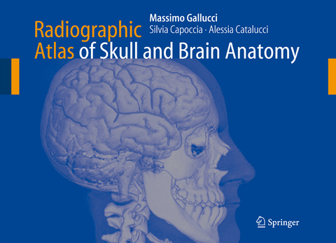
Radiographic Atlas of Skull and Brain Anatomy
Springer Berlin (Verlag)
978-3-642-07059-4 (ISBN)
- Keine Verlagsinformationen verfügbar
- Artikel merken
The book is very innovative, offering a detailed, original representation of normal anatomy of the skull and brain with more than 892 figures in black and white and colors. Plan X Ray, high resolution CT and MRI cuts on conventional planes are collected together with tridimensional reformatted images and with functional studies. Brain, skull, ear, orbit and vascular anatomy are treated in the same book, following systematic designs rather than technique dependent representations. High quality images are supported with extreme care in detail labeling and with color drawings and schemes. The book is geared toward neuroradiologists, radiologists, neurosurgeons, neurologists, anatomists.
Surface Anatomy.- Sectional Anatomy of the Telencephalon.- Brainstem and Cerebellum.- Cranial Nerves and Related Systems.- Functional Systems.- Vascular Anatomy.
From the reviews:
"I received directly from Massimo Gallucci his new book on brain anatomy. ... A text I loved as I love anatomy, the wonderful geography of the human body. ... work is really remarkable and deserve the maximum respect. ... I want to underline Massimo's presentation and dedication: it is of special interest as an opening to his rich and warm personality. A wonderful book, to have, to use, and to keep protected." (The Neuroradiology Journal, Vol. 21 (1), 2008)
| Erscheint lt. Verlag | 19.12.2010 |
|---|---|
| Zusatzinfo | X, 362 p. |
| Verlagsort | Berlin |
| Sprache | englisch |
| Maße | 279 x 210 mm |
| Gewicht | 1860 g |
| Themenwelt | Medizinische Fachgebiete ► Radiologie / Bildgebende Verfahren ► Radiologie |
| Schlagworte | anatomy • brain • Brainstem • Computed tomography (CT) • Neuroradiology • Radiology • Skull |
| ISBN-10 | 3-642-07059-0 / 3642070590 |
| ISBN-13 | 978-3-642-07059-4 / 9783642070594 |
| Zustand | Neuware |
| Haben Sie eine Frage zum Produkt? |
aus dem Bereich


