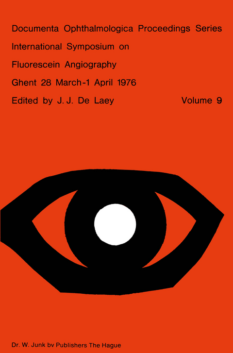
International Symposium on Fluorescein Angiography Ghent 28 March-1 April 1976
Kluwer Academic Publishers (Verlag)
978-90-6193-149-2 (ISBN)
This volume contains the papers presented at the International Symposium on Fluorescein Angiography held in Ghent, from 28 march to 1 april 1976, under the presidency of Prof. J. Fran90is. The book has been divided in several chapters corresponding to the sessions of the meeting. The same order has been followed as for the pre sentation of the papers. The discussions, however, immediately follow the papers concerned. During the meeting complications of fluorescein angio graphy have been discussed; this part will be presented as a separate chapter at the end of the volume. I wish to express my gratitude to all who contributed to this volume and to all the participants of ISF A-Ghent. I acknowledge also the cooperation of the publishers Dr. W. Junk, B.V. J.J. De Laey, M.D. XI EDITORIAL We must be respectfully grateful to Her Majesty the Queen, who very kindly extended her high patronage to the International Symposium on Fluorescein Angiography.
Session I — Instrumentation and technique.- Photography with corneal contact fundus cameras.- Clinical trials with the ‘Equator-Plus’ camera.- High speed fluorography.- Advances in TV-fluorangiography.- Improved interference filters for fluorescein angiography.- Fluorescein cycloscopy.- A new, TV-guided fundus camera.- Circulation parameters: comparison of both eyes by simultaneous fluorescein angiography.- Five years experience with automated processing for fluorescein angiography.- Angioscopy and colour angiography.- High speed human choroidal angiography using indocyanine green dye and a continuous light source.- Cine angiographic inflow measurements using fluorescein and indocyanine green.- Riboflavin fluorescence angiography.- Experimental angiography combined with routine black-and-white angiography made possible by utilizing a double-camera ‘MADO’head.- Illumination thresholds.- Demonstration of aqueous outflow by fluorescein injection into the anterior chamber after various types of glaucoma operations.- Session II — Retina I.- Fundamental aspects of posterior ocular circulation.- Quantitative aspects of fluorescein angiography.- Arteriovenous mean circulation time in the human retina.- The computerized elaboration of fluorangiographic data on retinal vascularization.- Television photometric technique for recording fluorescein dilution curves (dromofluorograms).- The Patho-Physiology of Retinal Vein Occlusion.- Natural Course and Classification of Patients with branch retinal vein obstruction.- Cotton-wool spots in retinal vein thrombosis.- Prognostic significance of fluorescein angiography in central retinal vein occlusion.- Traitement des occlusions veineuses rétiniennes.- Maculopathy and visual prognosis in retinal vein occlusion.- A comparativestudy of treated and non-treated cases of central retinal vein occlusion.- Surgery for vascular obstructions of the retina (Posada’s technique). F.A. aspects.- Session III — Choroidal circulation.- Anatomical correlation of the normal fluoroangiography of the fundus.- The development of the choroidal vascular system.- Fluorescein angiography and angio-architecture of the choroid.- Physiological anatomy of the choroidal vasculature.- Choroidal arterial occlusive disorders.- Etude de la circulation choriocapillaire du fond d’oeil humain.- Occlusion des veines choroidiennes.- Clinical application of indo-cyanine green angiography.- Watershed zone degeneration, a clinical syndrome?.- Consideration of the cilioretinal circulation.- Session IV — Choroid II.- Choroidal naevus and melanoma.- An angiographic and histopathologic confrontation concerning the chorio-retinal changes in front of a human malignant melanoma of the choroid.- On the significance of the bright dot-like fluorescence at different malignant intraocular tumorous growth.- Angiographic follow-up of choroidal melanoma.- Nodular choroidal masses in patients with sarcoidosis.- Optic disc and peripapillary choroid. A cinefluoroangiographic study.- A study of the optic disc fluorescence by photographic subtraction.- In vivo measurements of diffusion of fluorescein into the human optic nerve tissue.- Choroidal circulation in glaucoma.- The precursors of disci-form macular degeneration.- Differential perimetric profiles in disciform macular degeneration: stages of development.- Fluorangiographic study of chorioretinal lesions in high myopia.- Juvenile juxta-papillary hemorrhagic choroiditis.- Session V — Pigment epithelium and choroid.- Morphology of the pigment epithelium.- Bruch’s membrane.- Correlationof fluorescein angiography and histopathology.- Diseases affecting the pigment epithelium.- Acute multifocal posterior placoid pigment epitheliopathy and argon laser photocoagulation. An angiographic comparison.- Various presentations of pigment epitheliopathies and choriocapillaropathies.- Inflammations of the choroid.- Fluorescein Angiography in uveal effusion.- Angiographic fluorescéinique du fond d’oeil au cours d’une ophtalmie sympathique.- Lésions Chorioépithéliales initiales dans l’onchocercose oculaire.- Dominant Macular Dystrophies: Cystoid macular edema and butterfly dystrophy.- Sector refinitis pigmentosa with chronic disc edema.- Leakage from retinal capillaries in hereditary dystrophies.- Epithelium pigmentaire dans les hérédo-dégénérescences choriorétiniennes à prédominance choroidienne.- Fluorographic aspects of Stargardt’s disease.- Le signe du silence choroïdien dans les dégénérescences tapétoré tiniennes postérieures.- Session VI — Retina II.- Iris angiography in vascular diseases of the fundus.- Alteration in blood flow in the pathogenesis of diabetic retinopathy.- First lesions in infantile diabetic retinopathy. Angiofluoresceinic study.- Cystoid macular oedema in diabetic retinopathy.- Evaluation of diabetic retinopathy.- A model to quantitatively predict the course of diabetic retinopathy.- Réactions vasculaires et tissulaires de l’oeil provoquées par la cryocautérisation et par la coagulation au laser. Etude expérimentale à fluorescence.- Comparative study of retinal micro-aneurysm.- Fluorescein angiographic patterns of retinal arterial aneurysms.- Retinal vascular changes in Takayasu’s Disease (Pulseless Disease), occurrence and evolution of the lesion.- Retinopathy due to chronic carbon disulfidepoisoning.- Fluorescein angiography studies of posterior pole preretinal fibrosis (Macular Pucker).- Angiographic considerations about the preretinal membrane.- Session VII — Retina III.- Fluorography of the fundus periphery with rhegmatogenous retinal detachment.- Peripheral vascular diseases.- The value of fluorescein angiography in sickle cell retinopathy.- Fluorescein angiography in retrolental fibroplasia.- Alteration of retinal hemodynamics in retrolental fibroplasia.- Equatorial degenerations with atypical vessels.- Angiomatosis retinae.- Discussion on complications.- General review.
| Erscheint lt. Verlag | 30.6.1976 |
|---|---|
| Reihe/Serie | Documenta Ophthalmologica Proceedings Series ; 9 |
| Zusatzinfo | 367 Illustrations, black and white; 653 p. 367 illus. |
| Verlagsort | Dordrecht |
| Sprache | englisch |
| Maße | 155 x 235 mm |
| Themenwelt | Medizin / Pharmazie ► Medizinische Fachgebiete ► Augenheilkunde |
| ISBN-10 | 90-6193-149-5 / 9061931495 |
| ISBN-13 | 978-90-6193-149-2 / 9789061931492 |
| Zustand | Neuware |
| Haben Sie eine Frage zum Produkt? |
aus dem Bereich


