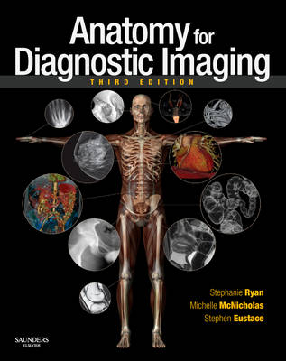
Anatomy for Diagnostic Imaging
Seiten
2010
|
3rd edition
W B Saunders Co Ltd (Verlag)
978-0-7020-2971-4 (ISBN)
W B Saunders Co Ltd (Verlag)
978-0-7020-2971-4 (ISBN)
- Titel erscheint in neuer Auflage
- Artikel merken
Zu diesem Artikel existiert eine Nachauflage
Addresses the anatomy of the imaging techniques including three-dimensional CT, cardiac CT, and CT and MR angiography as well as the anatomy of therapeutic interventional radiological techniques guided by fluoroscopy, ultrasound, CT and MR. This book is suitable for those training in radiology and preparing for the FRCR examinations.
This book covers the normal anatomy of the human body as seen in the entire gamut of medical imaging. It does so by an initial traditional anatomical description of each organ or system followed by the radiological anatomy of that part of the body using all the relevant imaging modalities. The third edition addresses the anatomy of new imaging techniques including three-dimensional CT, cardiac CT, and CT and MR angiography as well as the anatomy of therapeutic interventional radiological techniques guided by fluoroscopy, ultrasound, CT and MR. The text has been completely revised and over 140 new images, including some in colour, have been added. A series of 'imaging pearls' have been included with most sections to emphasise clinically and radiologically important points. The book is primarily aimed at those training in radiology and preparing for the FRCR examinations, but will be of use to all radiologists and radiographers both in training and in practice, and to medical students, physicians and surgeons and all who use imaging as a vital part of patient care. The third edition brings the basics of radiological anatomy to a new generation of radiologists in an ever-changing world of imaging.This book covers the normal anatomy of the human body as seen in the entire gamut of medical imaging. It does so by an initial traditional anatomical description of each organ or system followed by the radiological anatomy of that part of the body using all the relevant imaging modalities. The third edition addresses the anatomy of new imaging techniques including three-dimensional CT, cardiac CT, and CT and MR angiography as well as the anatomy of therapeutic interventional radiological techniques guided by fluoroscopy, ultrasound, CT and MR. The text has been completely revised and over 140 new images, including some in colour, have been added. A series of 'imaging pearls' have been included with most sections to emphasise clinically and radiologically important points. The book is primarily aimed at those training in radiology, but will be of use to all radiologists and radiographers both in training and in practice, and to medical students, physicians and surgeons and all who use imaging as a vital part of patient care. The third edition brings the basics of radiological anatomy to a new generation of radiologists in an ever-changing world of imaging.
Anatomy of new radiological techniques and anatomy relevant to new staging or treatment regimens is emphasised.
'Imaging Pearls' that emphasise clinically and radiologically important points have been added throughout.
The text has been revised to reflect advances in imaging since previous edition.
Over 100 additional images have been added.
Head and neck. Central nervous system. Spinal column and its contents. The thorax. The abdomen. The pelvis. The upper limb. The lower limb. The breast.
| Erscheint lt. Verlag | 24.9.2010 |
|---|---|
| Zusatzinfo | 520 illustrations (3 in full color); Illustrations |
| Verlagsort | London |
| Sprache | englisch |
| Gewicht | 1000 g |
| Themenwelt | Medizinische Fachgebiete ► Radiologie / Bildgebende Verfahren ► Radiologie |
| Studium ► 1. Studienabschnitt (Vorklinik) ► Anatomie / Neuroanatomie | |
| ISBN-10 | 0-7020-2971-8 / 0702029718 |
| ISBN-13 | 978-0-7020-2971-4 / 9780702029714 |
| Zustand | Neuware |
| Informationen gemäß Produktsicherheitsverordnung (GPSR) | |
| Haben Sie eine Frage zum Produkt? |
Mehr entdecken
aus dem Bereich
aus dem Bereich
Buch (2023)
Thieme (Verlag)
CHF 265,95


