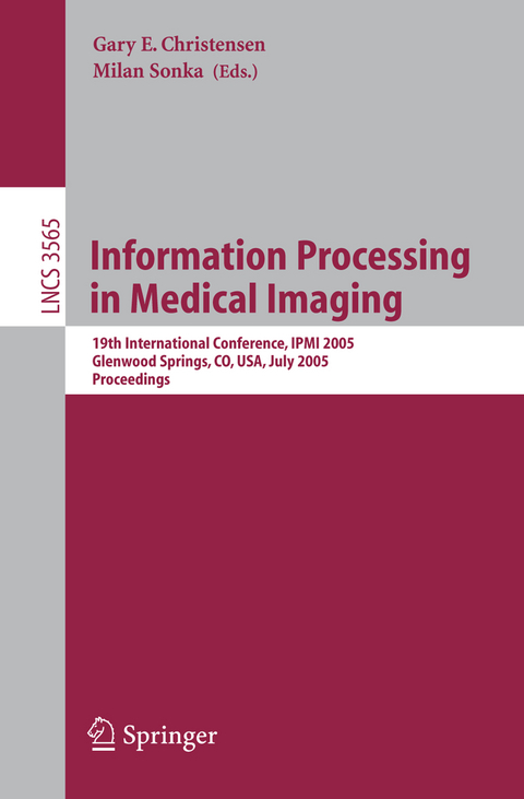
Information Processing in Medical Imaging
Springer Berlin (Verlag)
978-3-540-26545-0 (ISBN)
Shape and Population Modeling.- A Unified Information-Theoretic Approach to Groupwise Non-rigid Registration and Model Building.- Hypothesis Testing with Nonlinear Shape Models.- Extrapolation of Sparse Tensor Fields: Application to the Modeling of Brain Variability.- Bayesian Population Modeling of Effective Connectivity..- Diffusion Tensor Imaging and Functional Magnetic Resonance.- Fiber Tracking in q-Ball Fields Using Regularized Particle Trajectories.- Approximating Anatomical Brain Connectivity with Diffusion Tensor MRI Using Kernel-Based Diffusion Simulations.- Maximum Entropy Spherical Deconvolution for Diffusion MRI.- From Spatial Regularization to Anatomical Priors in fMRI Analysis.- Segmentation and Filtering.- CLASSIC: Consistent Longitudinal Alignment and Segmentation for Serial Image Computing.- Robust Active Appearance Model Matching.- Simultaneous Segmentation and Registration of Contrast-Enhanced Breast MRI.- Multiscale Vessel Enhancing Diffusion in CT Angiography Noise Filtering.- Poster Session 1.- Information Fusion in Biomedical Image Analysis: Combination of Data vs. Combination of Interpretations.- Parametric Medial Shape Representation in 3-D via the Poisson Partial Differential Equation with Non-linear Boundary Conditions.- Diffeomorphic Nonlinear Transformations: A Local Parametric Approach for Image Registration.- A Framework for Registration, Statistical Characterization and Classification of Cortically Constrained Functional Imaging Data.- PET Image Reconstruction: A Robust State Space Approach.- Multi-dimensional Mutual Information Based Robust Image Registration Using Maximum Distance-Gradient-Magnitude.- Tissue Perfusion Diagnostic Classification Using a Spatio-temporal Analysis of Contrast Ultrasound Image Sequences.- Topology PreservingTissue Classification with Fast Marching and Topology Templates.- Apparent Diffusion Coefficient Approximation and Diffusion Anisotropy Characterization in DWI.- Linearization of Mammograms Using Parameters Derived from Noise Characteristics.- Knowledge-Driven Automated Detection of Pleural Plaques and Thickening in High Resolution CT of the Lung.- Fundamental Limits in 3D Landmark Localization.- Computational Elastography from Standard Ultrasound Image Sequences by Global Trust Region Optimization.- Representing Diffusion MRI in 5D for Segmentation of White Matter Tracts with a Level Set Method.- Automatic Prediction of Myocardial Contractility Improvement in Stress MRI Using Shape Morphometrics with Independent Component Analysis.- Brain Segmentation with Competitive Level Sets and Fuzzy Control.- Coupled Shape Distribution-Based Segmentation of Multiple Objects.- Partition-Based Extraction of Cerebral Arteries from CT Angiography with Emphasis on Adaptive Tracking.- Regional Whole Body Fat Quantification in Mice.- Surface Matching via Currents.- A Genetic Algorithm for the Topology Correction of Cortical Surfaces.- Simultaneous Segmentation of Multiple Closed Surfaces Using Optimal Graph Searching.- A Generalized Level Set Formulation of the Mumford-Shah Functional for Brain MR Image Segmentation.- Integrable Pressure Gradients via Harmonics-Based Orthogonal Projection.- Design of Robust Vascular Tree Matching: Validation on Liver.- A Novel Parametric Method for Non-rigid Image Registration.- Transitive Inverse-Consistent Manifold Registration.- Cortical Surface Alignment Using Geometry Driven Multispectral Optical Flow.- Inverse Consistent Mapping in 3D Deformable Image Registration: Its Construction and Statistical Properties.- Poster Session 2.- Robust Nonrigid Multimodal Image Registration Using Local Frequency Maps.- Imaging Tumor Microenvironment with Ultrasound.- PDE-Based Three Dimensional Path Planning for Virtual Endoscopy.- Elastic Shape Models for Interpolations of Curves in Image Sequences.- Segmenting and Tracking the Left Ventricle by Learning the Dynamics in Cardiac Images.- 3D Active Shape Models Using Gradient Descent Optimization of Description Length.- Capturing Anatomical Shape Variability Using B-Spline Registration.- A Riemannian Approach to Diffusion Tensor Images Segmentation.- Coil Sensitivity Estimation for Optimal SNR Reconstruction and Intensity Inhomogeneity Correction in Phased Array MR Imaging.- Many Heads Are Better Than One: Jointly Removing Bias from Multiple MRIs Using Nonparametric Maximum Likelihood.- Unified Statistical Approach to Cortical Thickness Analysis.- ZHARP: Three-Dimensional Motion Tracking from a Single Image Plane.- Analysis of Event-Related fMRI Data Using Diffusion Maps.- Automated Detection of Small-Size Pulmonary Nodules Based on Helical CT Images.- Nonparametric Neighborhood Statistics for MRI Denoising.- Construction and Validation of Mean Shape Atlas Templates for Atlas-Based Brain Image Segmentation.- Multi-figure Anatomical Objects for Shape Statistics.- The Role of Non-Overlap in Image Registration.- Multimodality Image Registration Using an Extensible Information Metric and High Dimensional Histogramming.- Spherical Navigator Registration Using Harmonic Analysis for Prospective Motion Correction.- Tunneling Descent Level Set Segmentation of Ultrasound Images.- Multi-object Segmentation Using Shape Particles.
| Erscheint lt. Verlag | 24.6.2005 |
|---|---|
| Reihe/Serie | Image Processing, Computer Vision, Pattern Recognition, and Graphics | Lecture Notes in Computer Science |
| Zusatzinfo | XXI, 777 p. |
| Verlagsort | Berlin |
| Sprache | englisch |
| Maße | 155 x 235 mm |
| Gewicht | 1198 g |
| Themenwelt | Informatik ► Grafik / Design ► Digitale Bildverarbeitung |
| Schlagworte | Active appearance model • Active Shape Model • Bildgebende Verfahren (Medizin) • brain imaging • Cardiac Imaging • Computed tomography (CT) • fmri analysis • Image Analysis • Interpolation • Magnetic Resonance Imaging • Medical Image Computing • Medical Image Processing • medical image registration • Medical Image Sequences • Medical Imaging • MRI • Radiology • tissue |
| ISBN-10 | 3-540-26545-7 / 3540265457 |
| ISBN-13 | 978-3-540-26545-0 / 9783540265450 |
| Zustand | Neuware |
| Informationen gemäß Produktsicherheitsverordnung (GPSR) | |
| Haben Sie eine Frage zum Produkt? |
aus dem Bereich


