Head and Neck
Seiten
2009
Cambridge University Press
978-0-521-87999-6 (ISBN)
Cambridge University Press
978-0-521-87999-6 (ISBN)
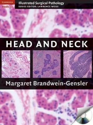
From the Cambridge Illustrated Surgical Pathology series this book comprehensively covers all of the methods utilized by pathologists to accurately diagnose diseases affecting all organs in the head and neck region. This full-color atlas is illustrated with more than 300 color photomicrographs and accompanied by a CD-ROM of all images in downloadable format.
Much of the diversity of head and neck diseases is due to the large number and broad function of the organs within this region. The surgical pathologist must become proficient in this subspecialty area in order to identify and categorize many different subtypes of lesions and diseases, including those affecting the thyroid and salivary glands. This book from the Cambridge Illustrated Surgical Pathology series comprehensively covers all of the methods utilized by pathologists to accurately diagnose diseases affecting all organs in the head and neck region. Coverage is not limited to findings from the light microscope but also includes other genetic, molecular, and immunologic diagnostic modalities, and the unique orientation allows the reader to follow the progression of disease states from incipient to advanced. This book is illustrated with more than 300 color photomicrographs and accompanied by a CD-ROM of all images in downloadable format.
Much of the diversity of head and neck diseases is due to the large number and broad function of the organs within this region. The surgical pathologist must become proficient in this subspecialty area in order to identify and categorize many different subtypes of lesions and diseases, including those affecting the thyroid and salivary glands. This book from the Cambridge Illustrated Surgical Pathology series comprehensively covers all of the methods utilized by pathologists to accurately diagnose diseases affecting all organs in the head and neck region. Coverage is not limited to findings from the light microscope but also includes other genetic, molecular, and immunologic diagnostic modalities, and the unique orientation allows the reader to follow the progression of disease states from incipient to advanced. This book is illustrated with more than 300 color photomicrographs and accompanied by a CD-ROM of all images in downloadable format.
Margaret Brandwein-Gensler, MD, is Director of Head and Neck Pathology, and Professor of Pathology and Otorhinolaryngology at Albert Einstein College of Medicine of Yeshiva University, Bronx, New York.
1. Sinonasal tract; 2. Nasopharynx; 3. Oral cavity; 4. Larynx; 5. Salivary glands; 6. Thyroid and parathyroid glands; 7. Jaws.
| Erscheint lt. Verlag | 12.10.2009 |
|---|---|
| Reihe/Serie | Cambridge Illustrated Surgical Pathology |
| Zusatzinfo | 33 Tables, unspecified; 538 Plates, color; 17 Halftones, unspecified |
| Verlagsort | Cambridge |
| Sprache | englisch |
| Maße | 220 x 285 mm |
| Gewicht | 2790 g |
| Themenwelt | Studium ► 2. Studienabschnitt (Klinik) ► Pathologie |
| ISBN-10 | 0-521-87999-X / 052187999X |
| ISBN-13 | 978-0-521-87999-6 / 9780521879996 |
| Zustand | Neuware |
| Haben Sie eine Frage zum Produkt? |
Mehr entdecken
aus dem Bereich
aus dem Bereich
Media-Kombination (2022)
Wolters Kluwer Health
CHF 669,95
Media-Kombination (2023)
Cambridge University Press
CHF 259,95

