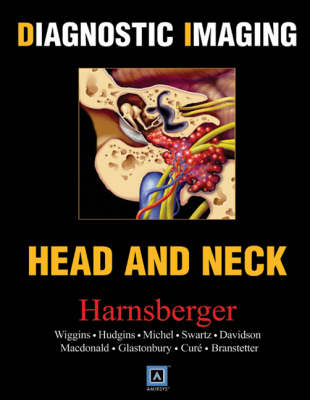
Head and Neck
Top 250 Diagnoses
Seiten
2004
W B Saunders Co Ltd (Verlag)
978-0-7216-2890-5 (ISBN)
W B Saunders Co Ltd (Verlag)
978-0-7216-2890-5 (ISBN)
- Titel ist leider vergriffen;
keine Neuauflage - Artikel merken
Part of the "Diagnostic Imaging" series, this work focuses on head and neck imaging. Every entity contains Clinical Presentation, Pathologic Features, Imaging Findings for the appropriate modalities, and Differential Diagnoses lists. Each chapter has anatomic drawings and gross and histologic pathology, and an image bank of case study variations.
Authored by a world-renowned team of researchers and teachers, this new text will become the outstanding one volume concise reference in Head and Neck Imaging. The third in our "Diagnostic Imaging" series, it has a templated, four-color format that makes finding information much easier. Each chapter has all the information you need to pinpoint a diagnosis. Every entity contains Clinical Presentation, Pathologic Features, Imaging Findings for the appropriate modalities, and Differential Diagnoses lists. In addition, each chapter has detailed anatomic drawings in full color and gross and histologic pathology, and an image bank of case study variations. There is no other "Head and Neck" text which is as comprehensive and user-friendly.
Authored by a world-renowned team of researchers and teachers, this new text will become the outstanding one volume concise reference in Head and Neck Imaging. The third in our "Diagnostic Imaging" series, it has a templated, four-color format that makes finding information much easier. Each chapter has all the information you need to pinpoint a diagnosis. Every entity contains Clinical Presentation, Pathologic Features, Imaging Findings for the appropriate modalities, and Differential Diagnoses lists. In addition, each chapter has detailed anatomic drawings in full color and gross and histologic pathology, and an image bank of case study variations. There is no other "Head and Neck" text which is as comprehensive and user-friendly.
The contents is divided into 15 sections, each with 16-25 major diagnoses in each section: 1.CPA-IAC 2.Skull Base 3.Temporal Bone 4.Orbit 5.Nose and Sinus 6.Pharyngeal Mucosal Space 7.Lymph Nodes 8.Larynx 9.Oral Cavity 10.Masticator Space 11.Parotid Space 12.Carotid Space 13.Midline Spaces 14.Visceral Space 15. Pediatric Lesions
| Erscheint lt. Verlag | 22.10.2004 |
|---|---|
| Reihe/Serie | Diagnostic Imaging |
| Zusatzinfo | Approx. 4400 illustrations |
| Verlagsort | London |
| Sprache | englisch |
| Maße | 216 x 279 mm |
| Gewicht | 3090 g |
| Themenwelt | Medizinische Fachgebiete ► Chirurgie ► Unfallchirurgie / Orthopädie |
| Medizin / Pharmazie ► Medizinische Fachgebiete ► HNO-Heilkunde | |
| Medizinische Fachgebiete ► Radiologie / Bildgebende Verfahren ► Radiologie | |
| ISBN-10 | 0-7216-2890-7 / 0721628907 |
| ISBN-13 | 978-0-7216-2890-5 / 9780721628905 |
| Zustand | Neuware |
| Haben Sie eine Frage zum Produkt? |
Mehr entdecken
aus dem Bereich
aus dem Bereich
Buch | Hardcover (2012)
Westermann Schulbuchverlag
CHF 44,90
Schulbuch Klassen 7/8 (G9)
Buch | Hardcover (2015)
Klett (Verlag)
CHF 29,90
Buch | Softcover (2004)
Cornelsen Verlag
CHF 23,90


