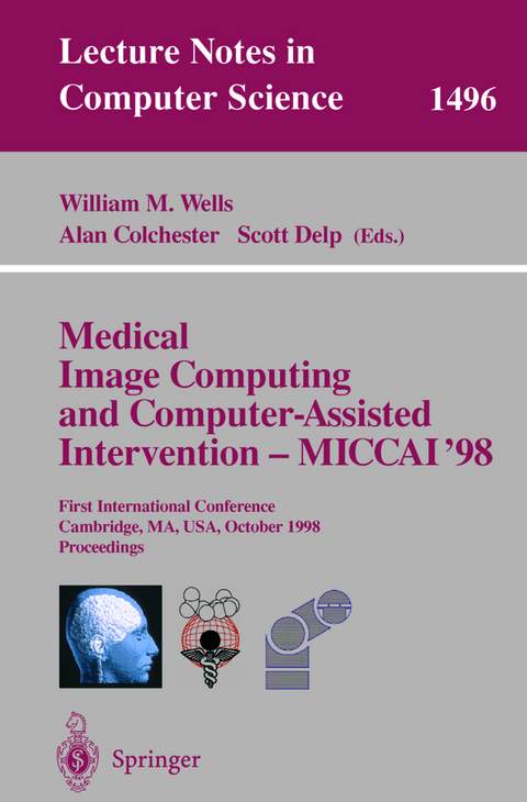
Medical Image Computing and Computer-Assisted Intervention - MICCAI'98
Springer Berlin
978-3-540-65136-9 (ISBN)
The 134 revised papers presented were carefully selected from a total of 243 submissions. The book is divided into topical sections on surgical planning, surgical navigation and measurements, cardiac image analysis, medical robotic systems, surgical systems and simulators, segmentation, computational neuroanatomy, biomechanics, detection in medical images, data acquisition and processing, neurosurgery and neuroscience, shape analysis, feature extraction, registration, and ultrasound.
Planning and evaluation of reorienting osteotomies of the proximal femur in cases of SCFE using virtual three-dimensional models.- Computer-aided planning of patellofemoral joint OA surgery: Developing physical models from patient MRI.- Computer assisted orthognathic surgery.- Computer-aided image-guided bone fracture surgery: Modeling, visualization, and preoperative planning.- A surgical planning and guidance system for high tibial osteotomies.- Measurement of intraoperative brain surface deformation under a craniotomy.- Clinical experience with a high precision image-guided neurosurgery system.- Three-dimensional reconstruction and surgical navigation in padiatric epilepsy surgery.- Treatment of pelvic ring fractures: Percutaneous computer assisted iliosacral screwing.- Model tags: Direct 3D tracking of heart wall motion from tagged MR images.- Quantitative three dimensional echocardiography: Methodology, validation, and clinical applications.- Measurement of 3D motion of myocardialmaterial points from explicit B-surface reconstruction of tagged MRI data.- 3D reconstruction from projection matrices in a C-arm based 3D-angiography system.- Multiscale vessel enhancement filtering.- Fast quantification of abdominal aortic aneurysms from CTA volumes.- 3-D fusion of biplane angiography and intravascular ultrasound for accurate visualization and volumetry.- Patient-specific analysis of left ventricular blood flow.- Dense 2D displacement reconstruction from SPAMM-MRI with constrained elastic splines: Implementation and validation.- Motion analysis of the right ventricle from MRI Images.- Magnetic resonance guided radiofrequency ablation: Creation and visualization of cardiac lesions.- Human versus robotic organ retraction during laparoscopic Nissen fundoplication.- A new laparoscope manipulator with an optical zoom.- A newly developed stereotactic robot with detachable drive for neurosurgery.- Calibration of video cameras to the coordinate system of a radiation therapytreatment machine.- An image overlay system for medical data visualization.- Volumetric image guidance via a stereotactic endoscope.- The application accuracy of the frameless implantable marker system and analysis of related affecting factors.- Multi-level strategy for Computer-Assisted Transbronchial Biopsy.- A fast, accurate and easy method to position oral implant using computed tomography.- Experimental protocol of accuracy evaluation of 6-D localizers for computer-integrated surgery: Application to four optical localizers.- Visualization and evaluation of prostate needle biopsy.- Virtual endoscope system with force sensation.- Using region-of-interest based finite element modelling for brain-surgery simulation.- An image processing environment for guiding vascular MR interventions.- Fluoroscopic image processing for computer-aided orthopaedic surgery.- Probe design to robustly locate anatomical features.- Concepts and results in the development of a hybrid tracking system for CAS.- Computer-assisted interstitial brachytherapy.- 3-D model supported prostate biopsy simulation and evaluation.- Human factors in tele-inspection and tele-surgery: Cooperative manipulation under asynchronous video and control feedback.- Computer assisted coronary intervention by use of on-line 3D reconstruction and optimal view strategy.- A robotic approach to HIFU based neurosurgery.- Virtual surgery system using deformable organ models and force feedback system with three fingers.- A modular surgical robotic system for image guided percutaneous procedures.- Optimum designed micro active forceps with built-in fiberscope for retinal microsurgery.- Gauging clinical practice: Surgical navigation for total hip replacement.- Adaptive template moderated spatially varying statistical classification.- Automatic quantification of MS lesions in 3D MRI brain data sets: Validation of INSECT.- Computer-aided diagnostic system for pulmonary nodules using helical CT images.- Enhanced spatial priors for segmentation of magnetic resonance imagery.- Exploring the discrimination power of the time domain for segmentation and characterization of lesions in serial MR data.- Reconstruction of the central layer of the human cerebral cortex from MR images.- Regularization of MR diffusion tensor maps for tracking brain white matter bundles.- Measurement of brain structures based on statistical and geometrical 3D segmentation.- Automatic identificaiton of cortical sulci using a 3D probabilistic atlas.- Segmentation and measurement of the cortex from 3D MR images.- A biomechanical model of soft tissue deformation, with applications to non-rigid registration of brain images with tumor pathology.- Building biomechanical models based on medical image data: An assessment of model accuracy.- Modelling of soft tissue deformation for laparoscopic surgery simulation.- A colour image processing method for melanoma detection.- Abnormal masses in mammograms: Detection using scale-orientation signatures.- Detecting and inferring brain activation from functional MRI by hypothesis-testing based on the likelihood ratio.- A fast technique for motion correction in DAS using a feature-based, irregular grid.- Autofocusing of clinical shoulder MR images for correction of motion artifacts.- Reconstruction of elasticity and attenuation maps in shear wave imaging: An inverse approach.- Understanding intensity non-uniformity in MRI.- An automatic threshold-based scaling method for enhancing the usefulness of Tc-HMPAO SPECT in the diagnosis of Alzheimer's disease.- Automatic computation of average brain models.- Brain shift modeling for use in neurosurgery.- Proximity constraints in deformable models for cortical surface identification.- Fast analysis of intracranical aneurysms based on interactive direct volume rendering and CTA.- Visualizing spatial resolution of linear estimation techniques of electromagnetic brain activity localization.- Biomechanical simulation of the vitreous humor in theeye using an Enhanced ChainMail Algorithm.- A biomechanical model of the human tongue and its clinical implications.- Three-dimensional joit kinematics using bone surface registration: A computer assisted approach with an application to the wrist joint in vivo.- Range of motion after total hip arthroplasty: Simulation of non-axisymmetric implants.- 4D shape-preserving modelling of bone growth.- AnatomyBrowser: A framework for integration of medical information.- Automatic, accurate surface model inference for dental CAD/CAM.- Initial in-vivo analysis of 3D heterogeneous brain computations for model-updated image-guided neurosurgery.- A new dynamic FEM-based subdivision surface model for shape recovery and tracking in medical images.- Automatic quantification of changes in the volume of brain structures.- Automatic analysis of normal brain dissymmetry of males and females in MR images.- Marching optimal-parameter ridges: An algorithm to extract shape loci in 3D images.- Singularities asfeatures of deformation grids.- Morphological analysis of terminal air spaces by means of micro-CT and confocal microscopy and simulation within a functional model of lung.- 2D+T acoustic boundary detection in echocardiography.- Automatically finding optimal working projections for the endovascular coiling of intracranical aneurysms.- Computer assisted quantitative analysis of deformities of the human spine.- Motion measurements in low-contrast X-ray imagery.- Pitfalls in comparing functional magnetic resonance imaging and invasive electrophysiology recordings.- Specification, modelling and visualization of arbitrarily shaped cut surfaces in the volume model.- An object-based volumetric deformable atlas for the improved localization of neuroanatomy in MR images.- Automated labeling of bronchial branches in virtual bronchoscopy system.- Building a complete surface model from sparse data using statistical shape models: Application to computer assisted knee surgery.- Constrained elasticsurface nets: Generating smooth surfaces from binary segmented data.- Assessing skill and learning in surgeons and medical students using a force feedback surgical simulator.- Virtual reality vitrectomy simulator.- An experimental image guided surgery simulator for hemicricolaryngectomy and reconstruction by tracheal autotransplantation.- Virtual endoscopy of mucin-producing pancreas tumors.- Augmented reality visualization for laparoscopic surgery.- Evaluation of control point selection in automatic, mutual information driven, 3D warping.- 3D/2D registration via skeletal near projective invariance in tubular objects.- Measuring global and local spatial correspondence using information theory.- Non-linear cerebral registration with sulcal constraints.- A double scanning procedure for visualisation of radiolucent objects in soft tissues: Application to oral implant surgery planning.- Interactive pre-operative selection of cutting constraints, and interactive force controlled knee surgery by a surgical robot.- Multimodal volume-based tumor neurosurgery planning in the virtual workbench.- Real-time tools for freehand 3D ultrasound.- Computer-based determination of the newborn's femoral head coverage using three-dimensional ultrasound scans.- Ultrasound imaging simulation: Application to the diagnosis of deep venous thromboses of lower limbs.- Isolating moving anatomy in ultrasound without anatomical knowledge: Application to computer-assisted pericardial punctures.- A new branching model: Application to carotid ultrasonic data.- Multi-modal volume registration using joint intensity distributions.- Multimodality deformable registration of pre- and intraoperative images for MRI-guided brain surgery.- A novel approach for the registration of 2D portal and 3D CT images for treatment setup verification in radiotherapy.- Multimodality imaging for epilepsy diagnosis and surgical focus localization: Three-dimensional image correlation and dual isotope SPECT.- Non-rigid multimodal image registration using mutual information.- Feature-based registration of medical images: Estimation and validation of the pose accuracy.- The correlation ratio as a new similarity measure for multimodal image registration.- Real-time registration of 3D cerebral vessels to X-ray angiograms.- Multi-object deformable templates dedicated to the segmentation of brain deep structures.- Non-rigid registration of breast MR images using mutual information.- A comparison of similarity measures for use in 2D-3D medical image registration.- Elastic model based non-rigid registration incorporating statistical shape information.- Image registration based on thin-plate splines and local estimates of anisotropic landmark localization uncertainties.- Segmentation of carpal bones from 3D CT images using skeletally coupled deformable models.- Segmentation of bone in clinical knee MRI using texture-based geodesic active contours.- Tensor controlled local structure enhancement of CT images for bonesegmentation.- Segmentation of magnetic resonance images using 3D deformable models.- Automatic segmentation of brain tissues and MR bias field correction using a digital brain atlas.- Robust brain segmentation using histogram scale-space analysis and mathematical morphology.- Vascular shape segmentation and structure extraction using a shape-based region-growing model.
| Erscheint lt. Verlag | 2.10.1998 |
|---|---|
| Reihe/Serie | Lecture Notes in Computer Science |
| Zusatzinfo | XLIV, 1258 p. 599 illus., 20 illus. in color. In 2 volumes, not available separately. |
| Verlagsort | Berlin |
| Sprache | englisch |
| Maße | 155 x 235 mm |
| Gewicht | 1572 g |
| Themenwelt | Mathematik / Informatik ► Informatik |
| Medizinische Fachgebiete ► Radiologie / Bildgebende Verfahren ► Radiologie | |
| Schlagworte | Algorithmen • Bildgebendes Verfahren • Bildverarbeitung • Biomechanics • Cardic Image Analysis • Computerunterstütztes Verfahren • Computer Version • Hardcover, Softcover / Informatik, EDV/Informatik • HC/Informatik, EDV/Informatik • Image Analysis • Maschinelles Sehen • Medical Robotic • Medizinische Informatik • Navigation • robot • Surgical Planning • Systems • Ultrasound |
| ISBN-10 | 3-540-65136-5 / 3540651365 |
| ISBN-13 | 978-3-540-65136-9 / 9783540651369 |
| Zustand | Neuware |
| Haben Sie eine Frage zum Produkt? |
aus dem Bereich

