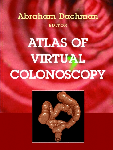
Atlas of Virtual Colonoscopy
Springer-Verlag New York Inc.
978-0-387-95511-7 (ISBN)
- Titel erscheint in neuer Auflage
- Artikel merken
Virtual colonoscopy is a rapidly developing technique that promises to be safer, more economical, and less intrusive than conventional diagnostic tests for colon cancer. This procedure allows the physician to look inside the body without having to insert a long tube into the colon, as in conventinal colonoscopy, or filling the colon with liquid barium, as with barium enema. VC has attracted the attention of the media and the public as a much-lauded alternative to current techniques. In response to this demand, renowned radiologist Dr. Abraham Dachman and a distinguished list of international contributors have put together a comprehensive atlas that depicts the entire range of this new technique. The illustrations are clear and true to life, showing normal and abnormal findings in virtual colonoscopy. This unique resource also points out critical clinical elements in using VC and summarizes the state of the art. The "Atlas of Virtual Colonoscopy" fills a void for all radiologists and gastroenterologists needing to or simply interested in learning about this exciting new technique.
Abraham H. Dachman, MD, is a nationally known specialist in abdominal imaging. He uses X-rays and advanced imaging equipment to visualize the structure and function of abdominal organs. This information is used to help diagnose disease, to assist in surgical planning, and to determine if treatments are effective. Dr. Dachman is known for his expertise in using computed tomography (CT scans) to create 3-D images of abdominal structures. This 3-D technology gives physicians an additional, valuable tool to better visualize tissue without performing an invasive procedure. He is a leading authority on virtual colonoscopy--using noninvasive CT technology to detect polyps and masses in the colon. In addition, he applies 3-D techniques to aid in the detection and staging of pancreatic cancer, and in the evaluation of tumor response to chemotherapy. An active researcher, Dr. Dachman has published several journal articles, book chapters, and books, including the first text on virtual colonoscopy, "The Atlas of Virtual Colonoscopy." In addition, he shares his knowledge about this emerging field through courses for radiologists who want to learn how to read virtual colonoscopy studies. He also has given presentations at dozens of scientific meetings around the United States. Andrea Laghi is Associate Professor in the Department of Radiological, Oncological and Pathological Sciences at the 'Sapienza' University of Rome and Director of the CT and MRI Unit at I.C.O.T. University Hospital, Latina, Italy. He currently chairs the Education Committee and is an Executive Committee member of the European Society of Gastrointestinal and Abdominal Radiology (ESGAR), and he is Coordinator of the ESGAR CT Colonography Hands-on Workshops. On behalf of ESGAR he is also member of the Education Committee of the United European Gastrointestinal Federation (UEGF). Professor Laghi is active member of the Associazione Italiana di Radiologia Medica, the European Society of Radiology (ESR) and the Radiological Society of North America (RSNA). He is currently a reviewer for several journals including European Radiology, Radiology, Investigative Radiology, The Lancet and Gastreonterology. Professor Laghi has been invited to present more than 200 lectures and is the author of over 300 published papers.
Part I - Text; 1. Virtual Colonoscopy: The Inside Story; 2. Background and Significance; 3. How Accurate is Virtual Colonoscopy?; 4. How to Perform and Interpret Virtual Colonoscopy; 5. Patient Preparation; 6. Innovative Display Techniques; 7. MRI; 8. Future Directions: Computer Aided Diagnosis; 9. A Word About Radiation Dose Part II - Atlas; 10. The Normal Colon; 11. Sessile Polyps; 12. Pedunculated Polyps; 13. Diminutive Lesion; 14. Flat Lesion; 15. Recognize Stool; 16. Large Masses and Post-Operative Colon; 17. Pitfalls and Artifacts; 18. IV Contrast; 19. Oral Contrast
| Erscheint lt. Verlag | 11.3.2003 |
|---|---|
| Vorwort | Joseph T. Ferrucci, J.H. Bond |
| Zusatzinfo | 451 black & white illustrations, 28 colour illustrations, 4 black & white tables |
| Verlagsort | New York, NY |
| Sprache | englisch |
| Maße | 216 x 279 mm |
| Gewicht | 1160 g |
| Einbandart | gebunden |
| Themenwelt | Medizinische Fachgebiete ► Innere Medizin ► Gastroenterologie |
| Medizin / Pharmazie ► Medizinische Fachgebiete ► Onkologie | |
| Medizin / Pharmazie ► Medizinische Fachgebiete ► Radiologie / Bildgebende Verfahren | |
| Studium ► 2. Studienabschnitt (Klinik) ► Anamnese / Körperliche Untersuchung | |
| ISBN-10 | 0-387-95511-9 / 0387955119 |
| ISBN-13 | 978-0-387-95511-7 / 9780387955117 |
| Zustand | Neuware |
| Haben Sie eine Frage zum Produkt? |
aus dem Bereich


