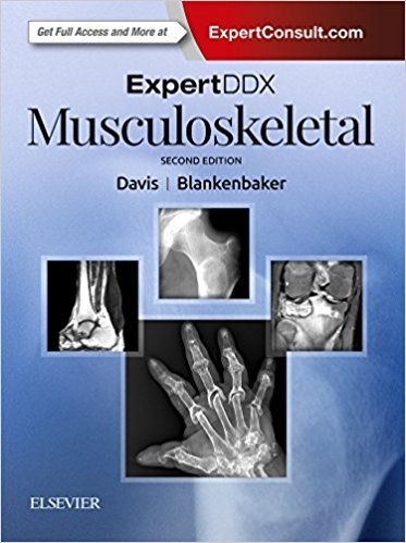
ExpertDDx: Musculoskeletal
Elsevier - Health Sciences Division (Verlag)
978-0-323-52483-4 (ISBN)
Zu diesem Buch erhalten Sie kostenlos ein eBook dazu.
More than 200 expert differential diagnosis lists based on imaging findings, clinical presentation, and anatomical location are organized according to likelihood of occurrence. Each includes at least eight clear, sharp, succinctly annotated images; a list of diagnostic possibilities sorted as common, less common, and rare but important; and brief, bulleted text offering helpful diagnostic clues.
- Includes all pertinent modalities-digital radiography, CT, MR, and ultrasound-focusing on quick reference for busy radiologists at the point of care
- Contains significantly revised content throughout, with many new examples of musculoskeletal conditions to help you refine your diagnoses
- Features new chapters on hypoechoic masses (ultrasound), hip impingement, and more, as well as new terminology, updated diagnostic facts, more ultrasound images, and new case examples in every chapter
- Covers hot topics such as FAI, subspinous impingement, ischiofemoral impingement, and iliopsoas impingement
- Expert ConsultT eBook version included with purchase. This enhanced eBook experience allows you to search all of the text, figures, Q&As, and references from the book on a variety of devices.
Kirkland W. Davis; Professor of Radiology, Musculoskeletal Imaging and Intervention, Department of Radiology, University of Wisconsin School of Medicine and Public Health, Madison, Wisconsin
Donna G Blankenbaker; Professor of Radiology, Musculoskeletal Imaging and Intervention, Department of Radiology, University of Wisconsin School of Medicine and Public Health, Madison, Wisconsin
ANATOMY BASED
Flat Bones
Flat Bones, Focally Expanded or Bubbly Lesion
Flat Bones, Permeative Lesion
Long Bone, Epiphyseal
Long Bone, Epiphyseal: Irregular or Stippled
Long Bone, Epiphyseal, Overgrowth/Ballooning
Long Bone, Epiphysis, Sclerosis/Ivory
Long Bone, Epiphyseal/Apophyseal/Subchondral Lytic Lesion
Long Bone, Metaphyseal
Long Bone, Metaphyseal Bands & Lines
Long Bone, Metaphyseal Cupping
Long Bone, Metaphyseal Fraying
Long Bone, Central Metaphyseal Lesion, Nonaggressive
Long Bone, Central Metaphyseal Lesion, Aggressive
Long Bone, Metaphyseal Lesion, Bubbly
Long Bone, Eccentric Metaphyseal Lesion, Nonaggressive
Long Bone, Eccentric Metaphyseal Lesion, Aggressive
Long Bone, Cortically Based Metaphyseal Lesion
Long Bone, Surface (Juxtacortical) Lesion
Long Bone, Metadiaphyseal
Long Bone, Central Diaphyseal Lesion, Nonaggressive
Long Bone, Diaphyseal Lesion, Aggressive: Adult
Long Bone, Diaphyseal Lesion, Aggressive: Child
Long Bone, Aggressive Diaphyseal Lesion With Endosteal Thickening
Long Bone, Cortically Based Diaphyseal Lesion, Sclerotic
Long Bone, Cortically Based Diaphyseal Lesion, Lytic
Long Bone, Diffuse Cortical/Endosteal Thickening
Tibial Metadiaphyseal Cortically Based Lesion
Long Bone, Undertubulation
Long Bone, Overtubulation
Lone Bone, Growth Plate
Growth Plate, Premature Physeal Closure
Growth Plate, Widened Physis
Periosteum
Periosteum: Aggressive Periostitis
Periosteum: Solid Periostitis
Periosteum: Bizarre Horizontal Periosteal Reaction
Periosteum: Periostitis Multiple Bones/Acropachy, Adult
Periosteum: Periostitis Multiple Bones, Child"
Joint Based
Arthritis With Normal Bone Density
Arthritis With Osteopenia
Arthritis With Productive Changes
Erosive Arthritis
Mixed Erosive/Productive Arthritis
Arthritis With Large Subchondral Cysts
Atrophic Joint Destruction
Arthritis Mutilans
Neuropathic Osteoarthropathy
Arthritis With Preserved Cartilage Space
Widened Joint Space
Ankylosis
Calcified Intraarticular Body/Bodies
Chondrocalcinosis
Periarticular Calcification
MCP-Predominant Arthritis
IP-Predominant Arthritis
Monoarthritis
Intraarticular Mass
Arthroplasty With Lytic/Cystic Lesions
Shoulder Girdle and Upper Arm
Clavicle Lesions, Nonarticular
Distal Clavicular Resorption
Proximal Humerus, Erosion Medial Metaphysis
Glenohumeral Malalignment
Anterosuperior Labral Variations/Pathology
Fluid Collections About the Shoulder
Elbow and Forearm
Radial Dysplasias/Aplasia
Forearm Deformity
Wrist and Hand
Carpal Cystic/Lytic Lesions
Abnormal Radiocarpal Angle
Fingers and Toes
Arachnodactyly
Soft Tissue Mass in Finger
Acro-osteolysis
Acro-osteosclerosis
Phalangeal Cystic/Lytic Lesions
Sesamoiditis
Short Metacarpal/Metatarsal
Ulnar Deviation (MCP Joints)
Swelling & Periostitis of Digit (Dactylitis)
Intevertebral Disc
Lesions Crossing Disc Space
Discal Mineralization
Paraspinal Abnormalities
Ossification/Calcification Anterior to C1
Paravertebral Ossification and Calcification
Linear Ossification Along Anterior Spine
Vertebral Shape
Bullet-Shaped Vertebra/Anterior Vertebral Body Beaking
Congenital & Acquired Childhood Platyspondyly
Fish (Biconcave) or H-Shaped Vertebra
Squaring of One or More Vertebra
Vertebral Lesions
Vertebral Body Sclerosis
Spinal Osteophytes
Lesions Originating in Vertebral Body
Lesions Originating in Posterior Elements
Ribs
Rib Notching, Inferior
Rib Notching, Superior
Solitary Rib Lesion
Pelvis
Sacroiliitis, Bilateral Symmetric
Sacroiliitis, Bilateral Asymmetric
Sacroiliitis, Unilateral
Symphysis Pubis With Productive Changes/Fusion
Symphysis Pubis, Widening
Supraacetabular Iliac Destruction
Hip and Thigh
Protrusio Acetabuli
Coxa Magna Deformity
Hip Labral Tears, Etiology
Knee and Lower Leg
Enlargement of Intercondylar Notch Distal Femur
Patellar Lytic Lesions
Tibial Bowing
Fluid Collections About the Knee
Popliteal Mass, Extraarticular
Alterations in Meniscal Size
Genu Valgum (Knock Knees)
Genu Varum (Bow Leg Deformity)
Foot and Ankle
Achilles Tendon Thickening/Enlargement
Calcaneal Erosions, Posterior Tubercle
Retrocalcaneal Bursitis
Soft Tissue Mass in Foot
Talar Beak
Tarsal Cystic/Lytic Lesions
IMAGE BASED View less >
Radiograph/CT, Osseous
Polyostotic Lesions, Adult
Polyostotic Lesions, Child
Solitary Geographic Lytic Lesions
Sclerotic Bone Lesion, Solitary
Sclerotic Bone Lesions, Multiple
Sclerotic Lesion With Central Lucency
Sequestration
Target Lesions of Bone
Matrix-Containing Bone Lesions
Benign Osseous Lesions That can Appear Aggressive
Metastases to Bone
Generalized Increased Bone Density, Adult
Generalized Increased Bone Density, Child
Sclerosing Dysplasias
Hypertrophic Callus Formation
Bone within Bone Appearance
Osteopenia
Osteoporosis, Generalized
Regional Osteoporosis
Cortical Tunneling
Pseudoarthrosis
Enthesopathy
Tendon & Ligamentous Ossification
Bone Age, Advanced
Bone Age, Delayed
Radiograph/CT, Soft Tissue
Soft Tissue Ossification
Nodular Calcification
Linear and Curvilinear Calcification
Soft Tissue Neoplasm Containing Calcification
MR, Osseous
Bone Marrow Edema Syndromes (Proximal Femur)
Subchondral Edema-Like Signal
Abnormal Epiphyseal Marrow Signal
Increased Marrow Fat
Marrow Hyperplasia
Bone Lesions With Fluid/Fluid Levels
MR, Soft Tissue
Lesion With Bright T1 Signal
Soft Tissue Lesion With Predominately Low T1 & T2 Signal
Soft Tissue Lesion With Fluid/Fluid Levels
Target Lesion of Soft Tissues
Cystic Mass
Subcutaneous Mass
Enlarged Muscle
Muscle Atrophy
Intermuscular Edema
Tenosynovitis/Tenosynovial Fluid
Enlarged Peripheral Nerves
MR, Joint
Intraarticular Low Signal Material, All Sequences
Ultrasound
Anechoic Mass
Hypoechoic Mass
Nuclear Medicine
Photopenic Lesions & False-Negative Scans
Soft Tissue Uptake on Bone Scan
Superscan
CLINICALLY BASED View less >
Shoulder and Upper Arm
Painful or Enlarged Sternoclavicular Joint
Rotator Cuff Symptoms
Shoulder Instability
Anteroinferior Labral/Capsule Injury
Nerve Entrapment, Shoulder
Elbow and Forearm
Elbow Deformities in Children and Young Adults
Lateral Elbow Pain
Medial Elbow Pain
Olecranon Bursitis
Nerve Entrapment, Elbow & Wrist
Wrist and Hand
Wrist Clicking/Clunking/Instability
Ulnar-Sided Wrist Pain
Radial-Sided Wrist Pain
Pelvis, Hip, and Thigh
Groin/Hip Pain
Lateral Hip Pain
Snapping Hip
Hip Impingement
Thigh Pain
Nerve Entrapment, Lower Extremity
Hip Pain, Elderly Patient
Painful Hip Replacement
Knee and Leg
Anterior Knee Pain
Medial Knee Pain
Calf Pain
Painful Knee Replacement
Ankle and Foot
Anterior Ankle Pain/Impingement
Medial Ankle Pain
Lateral Ankle Pain
Heel Pain
Pain in the Ball of the Foot
Pes Planovalgus (Flatfoot)
Cavus Foot Deformity
Congenital Foot Deformity
Diabetic Foot Complications
Spine
Painful Scoliosis
Systemic Disease
Arthritis in Teenager
Anemia With Musculoskeletal Manifestations
Osteonecrosis
Heterotopic Ossification
Rickets & Osteomalacia
Soft Tissue Contractures
Short Limb, Unilateral
Hemihypertrophy
Focal Gigantism/Macrodactyly
Dwarfism With Major Spine Involvement
Dwarfism With Short Extremities
Dwarfism With Short Ribs
Dwarfism With Horizontal Acetabular Roof
| Erscheinungsdatum | 19.12.2017 |
|---|---|
| Zusatzinfo | Approx. 3100 illustrations (3100 in full color) |
| Verlagsort | Philadelphia |
| Sprache | englisch |
| Maße | 222 x 281 mm |
| Gewicht | 2900 g |
| Einbandart | gebunden |
| Themenwelt | Medizinische Fachgebiete ► Chirurgie ► Unfallchirurgie / Orthopädie |
| Medizin / Pharmazie ► Medizinische Fachgebiete ► Orthopädie | |
| Medizinische Fachgebiete ► Radiologie / Bildgebende Verfahren ► Radiologie | |
| Schlagworte | muskuloskelettale Radiologie |
| ISBN-10 | 0-323-52483-4 / 0323524834 |
| ISBN-13 | 978-0-323-52483-4 / 9780323524834 |
| Zustand | Neuware |
| Haben Sie eine Frage zum Produkt? |
aus dem Bereich


