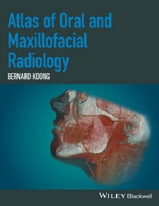
Atlas of Oral and Maxillofacial Radiology
John Wiley & Sons Inc (Verlag)
978-1-118-93964-2 (ISBN)
- Titel z.Zt. nicht lieferbar
- Versandkostenfrei
- Auch auf Rechnung
- Artikel merken
Focusing on the essentials of radiologic interpretation, this is a go-to companion for clinicians in everyday practice who have radiologically identified a potential abnormality, as well as a comprehensive study guide for students at all levels of dentistry, surgery and radiology.
- Unique lesion-based problem solving chapter makes this an easy-to-use reference in a clinical setting
- Includes 2D intraoral radiography, the panoramic radiograph, cone beam CT, multidetector CT and MRI
- Multiple cases are presented in order to demonstrate the variation in the radiological appearances of conditions affecting the jaws and teeth
- Special focus on conditions where diagnostic imaging may substantially contribute to diagnosis
Bernard Koong is a highly experienced oral and maxillofacial radiologist practicing full time multimodality clinical radiology. He is a founding partner of Envision Medical Imaging, a multidisciplinary fully comprehensive private radiology group in Australia and also consults internationally. He has personally reported over 200,000 radiological studies involving a wide variety of imaging techniques. Having completed his specialist training in oral and maxillofacial radiology at the University of Toronto, Bernard now holds the position of Clinical Professor at the University of Western Australia, where he coordinates and delivers the oral and maxillofacial radiology lectures for the undergraduate and postgraduate courses. He also has a long history of providing oral and maxillofacial radiology courses for other universities as well as surgery and radiology programmes across Australasia. As an invited speaker, Bernard has presented more than 100 lectures to the dental and medical professions internationally, and is a member of the Editorial Board of Clinical Oral Implant Research.
List of Contributors, xi
Preface, xii
Acknowledgements, xiii
How to Use This Atlas, xiv
1 Problem Solving Diagrams, 1
1.1 Opaque and largely opaque conditions related to the jaws, 1
Common conditions, 1
Less common conditions, 1
1.2 Lucent lesions of the jaws, 2
Common conditions, 2
Less common conditions, 2
1.3 Mixed density lesions of the jaws, 3
Common conditions, 3
Less common conditions, 3
2 Radiological Anatomy, 4
2.1 The panoramic radiograph, 4
2.2 Identification of teeth - FDI (Fédération Dentaire Internationale) World Dental Federation notation, 8
2.3 Cone beam computed tomography, 11
Axial, 11
Sagittal, 18
Coronal, 22
3 Anomalies Related to the Teeth, 28
3.1 Supernumerary teeth, 28
3.2 Congenital absence, 30
3.3 Delayed and early development/eruption, 31
3.4 Ectopic development and eruption, 32
3.5 Impaction, 36
3.6 Macrodontia, 40
3.7 Microdontia, 41
3.8 Dilaceration, 42
3.9 Enamel pearl, 42
3.10 Talon cusp, 43
3.11 Dens invaginatus, 44
3.12 Dens evaginatus, 45
3.13 Taurodontism, 45
3.14 Fusion, 46
3.15 Gemination, 47
3.16 Concrescence, 47
3.17 Amelogenesis imperfecta, 48
3.18 Dentinogenesis imperfecta, 49
3.19 Dentin dysplasia, 50
3.20 Secondary and tertiary dentin, 51
3.21 Pulp stones, 52
3.22 Hypercementosis, 53
4 Conditions Related to Loss of Tooth Structure, 54
4.1 Caries, 54
Interproximal caries, 54
Pit and fissure caries, 54
Root caries, 55
4.2 Attrition, 59
4.3 Abrasion, 60
4.4 Erosion, 61
4.5 Internal resorption, 61
4.6 External resorption, 62
4.7 Fracture related to trauma, 63
5 Inflammatory Lesions of the Jaws, 64
5.1 Periapical inflammatory lesions, 64
Post-treatment appearances of periapical lesions, 65
Re-establishment of normal periapical structures, 65
Variant trabecular architecture, 65
Fibrous healing, 65
Periapical osseous prominence at the maxillary sinus base, 66
5.2 Periodontal inflammatory disease, 74
5.3 Pericoronitis, 83
5.4 Osteomyelitis of the jaws, 86
5.5 Dentoalveolar and jaw infections involving the adjacent soft tissues, 88
6 Osteoradionecrosis and Osteonecrosis of the Jaws, 92
6.1 Osteoradionecrosis of the jaws, 92
6.2 Osteonecrosis of the jaws, 96
7 Hamartomatous/Hyperplastic Bony Opacities and Prominences Involving the Jaws, 97
7.1 Torus palatinus, 97
7.2 Torus mandibularis, 98
7.3 Exostoses, 100
7.4 Bone island, 101
8 Cysts and Cyst-like Lesions Involving the Jaws, 108
Odontogenic cysts and cyst -like lesions, 108
8.1 Radicular cyst, 108
8.2 Residual cyst, 114
8.3 Dentigerous cyst, 115
8.4 Buccal bifurcation cyst, 122
8.5 Keratocystic odontogenic tumour, 124
8.6 Basal cell naevus syndrome, 127
8.7 Lateral periodontal cyst, 128
8.8 Glandular odontogenic cyst, 130
Non-odontogenic cysts and cyst -like lesions, 130
8.9 Simple bone cyst, 130
8.10 Nasopalatine duct cyst, 136
8.11 Nasolabial cyst, 138
9 Fibro-osseous Lesions of the Jaws, 140
9.1 Fibrous dysplasia, 140
9.2 Cemento-osseous dysplasia, 145
9.3 Ossifying fibroma, 150
10 Benign Tumours Involving the Jaws, 153
ODONTOGENIC BENIGN TUMOURS, 153
10.1 Ameloblastoma, 153
10.2 Calcifying epithelial odontogenic tumour, 159
10.3 Odontoma, 160
10.4 Ameloblastic fibroma, 162
10.5 Ameloblastic fibro-odontoma, 163
10.6 Adenomatoid odontogenic tumour, 165
10.7 Calcifying cystic odontogenic tumour, 166
10.8 Odontogenic myxoma, 167
10.9 Cementoblastoma, 169
NON-ODONTOGENIC BENIGN TUMOURS INVOLVING THE JAWS, 170
10.10 Osteoma, 170
10.11 Gardner syndrome, 173
10.12 Osteochrondroma, 174
10.13 Schwannoma (within the jaws), 174
10.14 Osteoblastoma, 175
10.15 Osteoid osteoma, 176
10.16 Desmoplastic fibroma, 177
11 Malignant Tumours Involving the Jaws, 178
11.1 Imaging of malignancies involving the jaws, 178
11.2 Radiological features of malignancies involving the jaws, 178
11.3 Features of some malignancies which more commonly involve the jaws, 179
12 Vascular Anomalies of the Mid- and Lower Face, 191
VASCULAR TUMOURS (PROLIFERATIVE NEOPLASMS), 191
12.1 Haemangioma, 191
12.2 Other lesions included in this grouping, 193
VASCULAR MALFORMATIONS, 193
Complications, 193
12.3 Low-flow lesions, 193
Venolymphatic malformations or lymphangiomas, 193
Capillary malformations, 193
Venocavernous malformations, 194
12.4 High-flow lesions, 197
Arteriovenous malformations, 197
13 Other Diseases Affecting the Jaws, 199
13.1 Central giant cell granuloma, 199
13.2 Cherubism, 203
13.3 Aneurysmal bone cyst, 204
13.4 Langerhans cell histiocytosis, 205
13.5 Paget disease of bone, 208
14 Other Morphological Anomalies Involving the Jaws, 210
14.1 Hemimandibular hyperplasia, 210
14.2 Acromegaly, 212
14.3 Mandibular and hemimandibular hypoplasia, 212
14.4 Stafne defect, 214
14.5 Cleft lip and palate, 216
15 Other Systemic Disorders that may Involve the Jaws, 219
15.1 Osteopenic appearance of the jaws, 219
15.2 Increased density of the jaws, 221
15.3 Alterations in jaw size, 221
15.4 Changes to jaw morphology, 221
15.5 Dentoalveolar alterations, 221
16 Common Opacities in the Orofacial Soft Tissues, 222
16.1 Tonsillar calcifications, 222
16.2 Lymph node calcifications, 224
16.3 Stylohyoid ligamentous ossification, 225
16.4 Thyroid and triticeous cartilage calcifications, 226
16.5 Arterial calcifications related to arteriosclerosis, 228
16.6 Phlebolith, 231
16.7 Sialoliths, 231
16.8 Paranasal and nasal calcifications, 236
16.9 Myositis ossificans, 236
17 Trauma and Fractures, 238
TEETH AND SUPPORTING STRUCTURES, 238
17.1 Subluxation, 238
17.2 Luxation, 239
17.3 Avulsion, 240
17.4 Fracture of teeth, 241
FACIAL BONES, 245
17.5 Mandibular fractures, 245
17.6 N asal fracture, 247
17.7 Zygomaticomaxillary complex fracture, 248
17.8 Orbital blow-out fracture, 248
17.9 Le Fort fractures, 249
Le Fort I, 249
Le Fort II, 249
Le Fort III, 249
17.10 Other complex facial fractures, 249
18 Temporomandibular Joints, 250
18.1 Imaging the temporomandibular joints, 250
Panoramic radiograph, 250
Other plain film studies and dedicated conventional tomography, 250
Cone beam computed tomography (CBCT), 250
Multidetector (multislice) computed tomography (MDCT), 250
Magnetic resonance imaging (MRI), 250
18.2 Condylar hyperplasia, 250
18.3 Coronoid hyperplasia, 252
18.4 Condylar hypoplasia, 253
18.5 Bifid condyle, 255
18.6 Internal derangements of the temporomandibular joint, 256
18.7 Ganglion cysts, 261
18.8 Degenerative joint disease, 262
18.9 Inflammatory and erosive arthropathies, 268
18.10 Osteochrondroma, 270
18.11 Malignant tumours, 271
18.12 Synovial chondromatosis, 272
18.13 Calcium pyrophosphate deposition disease, 273
18.14 Ankylosis, 274
18.15 Other lesions affecting the temporomandibular joints, 275
18.16 Other non-temporomandibular joint conditions contributing to pain/dysfunction in the region of the temporomandibular joint and related structures, 275
19 Nasal Cavity, Paranasal Sinuses and Upper Aerodigestive Tract Impressions, 277
NASAL CAVITY AND PARANASAL SINUSES, 277
19.1 N ormal variations and developmental anomalies, 277
Variations in pneumatisation, 277
Accessory ethmoid air cells, 277
Aberrant transiting structures, 277
Accessory ostia, 277
Aberrant anatomical position, 277
Others, 277
19.2 Odontogenic conditions and dentoalveolar lesions, 280
19.3 Findings related to dental procedures, 280
Oroantral communication, 280
Tooth displacement, 280
Dental implants, 282
Periapical osseous healing, 282
19.4 Inflammatory paranasal sinus disease, 284
Acute rhinosinusitis, 284
Chronic rhinosinusitis, 286
Silent sinus syndrome, 287
Mucous retention cysts, 287
Sinonasal mucoceles, 288
Fungal rhinosinusitis, 289
Allergic fungal rhinosinusitis, 289
Sinonasal mycetoma, 290
Invasive fungal rhinosinusitis, 291
Sinonasal polyposis, 292
Antrochoanal polyps, 293
Granulomatous sinonasal inflammatory disease, 293
Granulomatosis with polyangiitis (previously known as Wegener granulomatosis), 294
Sarcoidosis, 294
Nasal cocaine necrosis, 295
19.5 Neoplastic disease, 296
Benign tumours, 296
Juvenile angiofibroma, 296
Sinus osteoma, 296
Sinonasal inverting papilloma, 297
Sinonasal cancers, 297
Sinonasal SCCa, 298
Sinonasal adenocarcinoma, 300
Minor salivary gland adenoid cystic carcinoma, 300
Sinonasal undifferentiated carcinoma, 300
Esthesioneuroblastoma or olfactory neuroblastoma, 301
Lymphoma, 302
PHARYNGEAL AIRWAY IMPRESSIONS, 303
19.6 Summary of causes of nasopharyngeal narrowing, 303
19.7 Summary of causes of oropharyngeal narrowing, 303
19.8 Malignant disease, 303
Nasopharyngeal carcinoma (NPC), 303
Oropharyngeal squamous cell carcinoma, 304
19.9 Benign entities, 305
Tornwald cyst, 305
Tortuous carotid arteries, 305
Lingual thyroid, 305
Foreign body ingestion, 307
19.10 Inflammatory lesions, 307
Tonsil hypertrophy and adenoid hypertrophy, 307
Retention cysts, 307
Tonsillitis, 308
Tonsillar and peritonsillar abscess, 309
Retropharyngeal space abscess, 310
Acute longus colli tendinitis, 310
19.11 Retropharyngeal adenopathy, 311
20 The Skull Base, 312
CONSTITUTIONAL AND DEVELOPMENTAL VARIATIONS, 312
20.1 Ossification of the interclinoid ligaments, 312
20.2 Benign notochordal cell tumour (ecchordosis physaliphora), 313
20.3 Persistence of the craniopharyngeal canal, 314
20.4 Arrested pneumatisation of the skull base, 315
20.5 Meningoencephaloceles, 316
20.6 Nasolacrimal duct mucocele (dacryocystocele), 317
20.7 Empty sella syndrome, 318
LESIONS OF THE SKULL BASE, 319
20.8 Pituitary macroadenoma, 319
20.9 Clival chordoma, 320
20.10 Skull base meningioma, 321
20.11 Skull base metastasis, 323
20.12 Chondrosarcoma, 324
20.13 Lymphoma, 325
20.14 Skull base plasmacytoma/multiple myeloma, 326
20.15 Langerhans cell histiocytosis, 327
20.16 Fibrous dysplasia, 327
20.17 Paget disease, 328
20.18 Petrous apex lesions, 329
EXPANSION OF SKULL BASE FORAMINA, 331
20.19 Nerve sheath tumours, 331
20.20 Perineural metastatic disease, 332
21 The Cervical Spine, 333
CONGENITAL VARIATIONS, 333
DEGENERATIVE DISEASE, 336
21.1 Cervical spondylosis, 336
21.2 Diffuse idiopathic hyperostosis, 337
21.3 Ossification of the posterior longitudinal ligament, 338
INFLAMMATORY AND DEPOSITIONAL CONDITIONS, 339
21.4 Rheumatoid arthritis, 339
21.5 Ankylosing spondylitis, 340
21.6 Osteomyelitis/discitis/facetal septic arthritis, including tuberculosis, 341
TUMOURS AND TUMOUR-LIKE LESIONS, 342
21.7 Metastatic tumours, 342
21.8 Multiple myeloma, 344
21.9 Aneurysmal bone cysts, 344
21.10 Peripheral nerve sheath tumours, 345
Index, 347
| Erscheinungsdatum | 20.03.2017 |
|---|---|
| Verlagsort | New York |
| Sprache | englisch |
| Maße | 221 x 283 mm |
| Gewicht | 1274 g |
| Einbandart | gebunden |
| Themenwelt | Medizin / Pharmazie ► Medizinische Fachgebiete ► Chirurgie |
| Medizinische Fachgebiete ► Radiologie / Bildgebende Verfahren ► Radiologie | |
| Medizin / Pharmazie ► Zahnmedizin | |
| Schlagworte | zahnärztliche Radiologie • zahnärztliches Röntgen |
| ISBN-10 | 1-118-93964-6 / 1118939646 |
| ISBN-13 | 978-1-118-93964-2 / 9781118939642 |
| Zustand | Neuware |
| Haben Sie eine Frage zum Produkt? |
aus dem Bereich


