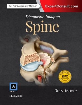
Diagnostic Imaging: Spine
Elsevier - Health Sciences Division (Verlag)
978-0-323-37705-8 (ISBN)
- Titel erscheint in neuer Auflage
- Artikel merken
Covers the latest advancements in imaging the postoperative spine, including bone morphogenetic protein (BMP) utilization
Includes additional genetic information, such as OMIM entry numbers, where appropriate
Highlights updates to new classification and grading schemes
Hundreds of full-color pathology images are carefully annotated to help pinpoint the most relevant factors
New references direct you to additional trustworthy resources
Bulleted lists provide guidance through the intricacies of the spine
Presents brand new images and cases to keep you at the forefront of your field
Expert Consult eBook version included with purchase. This enhanced eBook experience allows you to search all of the text, figures, and references from the book on a variety of devices
Jeffrey S. Ross, MD, is the returning co-lead for this edition. He is Professor of Radiology at Mayo Clinic College of Medicine and Science and practices neuroradiology at the Mayo Clinic. Ross was an AJNR Senior Editor from 2006-2015, is a member of the editorial board for 3 other journals, and a manuscript reviewer for 10 journals. He became Editor-in-Chief of the AJNR in July 2015. He received the Gold Medal Award from the ASSR in 2013. Kevin A. Moore, MD, is the returning co-lead for this edition. He is Adjunct Professor of Neuroradiology and Pediatric Radiology at Primary Children's Medical Center at the University of Utah, and is Board Certified by the American Board of Radiology in Diagnostic Radiology, Pediatric Radiology, and Neuroradiology.
Part I: Congenital and Genetic Disorders
SECTION 1: CONGENITAL
NORMAL ANATOMICAL VARIATIONS
Normal Anatomy
Measurement Techniques
MR Artifacts
Normal Variant
Craniovertebral Junction Variants
Ponticulus Posticus
Ossiculum Terminale
Conjoined Nerve Roots
Limbus Vertebra
Filum Terminale Fibrolipoma
Bone Island
Ventriculus Terminalis
CHIARI DISORDERS
Chiari 0
Chiari 1
Complex Chiari
Chiari 2
Chiari 3
ABNORMALITIES OF NEURULATION
Approach to Spine and Spinal Cord Development
Myelomeningocele
Lipomyelomeningocele
Lipoma
Dorsal Dermal Sinus
Simple Coccygeal Dimple
Dermoid Cysts
Epidermoid Cysts
ANOMALIES OF THE CAUDAL CELL MESS
Tethered Spinal Cord
Segmental Spinal Dysgenesis
Caudal Regression Syndrome
Terminal Myelocystocele
Anterior Sacral Meningocele
120 Sacral Extradural Arachnoid Cyst
Sacrococcygeal Teratoma
ANOMALIES OF NOTOCHORD AND VERTEBRAL FORMATION
Craniovertebral Junction Embryology
Paracondylar Process
Split Atlas
Klippel-Feil Spectrum
Failure of Vertebral Formation
Vertebral Segmentation Failure
Diastematomyelia
Partial Vertebral Duplication
Incomplete Fusion, Posterior Element
Neurenteric Cyst
DEVELOPMENTAL ABNORMALITIES
Os Odontoideum
Lateral Meningocele
Dorsal Spinal Meningocele
Dural Dysplasia
GENETIC DISORDERS
Neurofibromatosis Type 1
Neurofibromatosis Type 2
Achondroplasia
Mucopolysaccharidoses
Sickle Cell Disease
Osteogenesis Imperfecta
Tuberous Sclerosis
Osteopetrosis
Gaucher Disease
Ochronosis
Connective Tissue Disorders
Spondyloepiphyseal Dysplasia
Thanatophoric Dwarfism
SECTION 2: SCOLIOSIS AND KYPHOSIS
Introduction to Scoliosis
Scoliosis
Kyphosis
Degenerative Scoliosis
Flat Back Syndrome
Scoliosis Instrumentation
Part II: Trauma
SECTION 1: VERTEBRAL COLUMN, DISCS, AND PARASPINAL MUSCLE
Fracture Classification
Atlantooccipital Dislocation
Ligamentous Injury
Occipital Condyle Fracture
Jefferson C1 Fracture
Odontoid C2 Fracture
Burst C2 Fracture
Hangman's C2 Fracture
Apophyseal Ring Fracture
Cervical Hyperflexion Injury
Cervical Hyperextension Injury
Cervical Hyperextension-Rotation Injury
Cervical Burst Fracture
Cervical Hyperflexion-Rotation Injury
Cervical Lateral Flexion Injury
Cervical Posterior Column Injury
Traumatic Disc Herniation
Thoracic and Lumbar Burst Fracture
Facet-Lamina Thoracolumbar Fracture
Fracture Dislocation
Chance Fracture
Thoracic and Lumbar Hyperextension Injury
Anterior Compression Fracture
Lateral Compression Fracture
Lumbar Facet-Posterior Fracture
Sacral Traumatic Fracture
Pedicle Stress Fracture
Sacral Insufficiency Fracture
SECTION 2: CORD, DURA, AND VESSELS
SCIWORA
Post-traumatic Syrinx
Presyrinx Edema
Spinal Cord Contusion-Hematoma
Idiopathic Spinal Cord Herniation
Central Spinal Cord Syndrome
Traumatic Dural Tear
Traumatic Epidural Hematoma
Traumatic Subdural Hematoma
Vascular Injury, Cervical
Traumatic Arteriovenous Fistula
Wallerian Degeneration
Part III: Degenerative Diseases and Arthritides
SECTION 1: DEGENERATIVE DISEASES
Nomenclature of Degenerative Disc Disease
Degenerative Disc Disease
Degenerative Endplate Changes
Degenerative Arthritis of the CVJ
Disc Bulge
Anular Fissure, Intervertebral Disc
Cervical Intervertebral Disc Herniation
Thoracic Intervertebral Disc Herniation
Lumbar Intervertebral Disc Herniation
Intervertebral Disc Extrusion, Foraminal
Cervical Facet Arthropathy
Lumbar Facet Arthropathy
Facet Joint Synovial Cyst
Baastrup Disease
Bertolotti Syndrome
Schmorl Node
Scheuermann Disease
Acquired Lumbar Central Stenosis
Congenital Spinal Stenosis
Cervical Spondylosis
DISH
OPLL
Ossification Ligamentum Flavum
Periodontoid Pseudotumor
SECTION 2: SPONDYLOLISTHESIS AND SPONDYLOLYSIS
Spondylolisthesis
Spondylolysis
Instability
SECTION 3: INFLAMMATORY, CRYSTALLINE, AND MISCELLANEOUS ARTHRITIDES
Adult Rheumatoid Arthritis
Juvenile Idiopathic Arthritis
Spondyloarthropathy
Neurogenic (Charcot) Arthropathy
Hemodialysis Spondyloarthropathy
Ankylosing Spondylitis
CPPD
Gout
Longus Colli Calcific Tendinitis
Part IV: Infection and Inflammatory Disorders
SECTION 1: INFECTIONS
Pathways of Spread
Spinal Meningitis
Pyogenic Osteomyelitis
Tuberculous Osteomyelitis
Fungal and Miscellaneous Osteomyelitis
Osteomyelitis, C1-C2
Brucellar Spondylitis
Septic Facet Joint Arthritis
Paraspinal Abscess
Epidural Abscess
Subdural Abscess
Abscess, Spinal Cord
Viral Myelitis
HIV Myelitis
Syphilitic Myelitis
Opportunistic Infections
Echinococcus
Schistosomiasis
Cysticercosis
SECTION 2: INFLAMMATORY AND AUTOIMMUNE DISORDERS
Acute Transverse Myelopathy
Idiopathic Acute Transverse Myelitis
Multiple Sclerosis
Neuromyelitis Optica
ADEM
Guillain-Barré Syndrome
CIDP
Sarcoidosis
Subacute Combined Degeneration
HARDWARE
Metal Artifact
Occipitocervical Fixation
Plates and Screws
Cages
Interbody Fusion Devices
Interspinous Spacing Devices
Cervical Artificial Disc
Lumbar Artificial Disc
Hardware Failure
Bone Graft Complications
rhBMP-2 Complications
Heterotopic Bone Formation
Chronic Recurrent Multifocal Osteomyelitis
Grisel Syndrome
Paraneoplastic Myelopathy
Part V: Neoplasms, Cysts, and Other Masses
SECTION 1: NEOPLASMS
INTRODUCTION AND OVERVIEW
Spread of Neoplasms
EXTRADURAL
Blastic Osseous Metastases
Lytic Osseous Metastases
Hemangioma
Osteoid Osteoma
Osteoblastoma
Aneurysmal Bone Cyst
Giant Cell Tumor
Osteochondroma
Chondrosarcoma
Osteosarcoma
Chordoma
Ewing Sarcoma
Lymphoma
Leukemia
Plasmacytoma
Multiple Myeloma
Neuroblastic Tumor
Langerhans Cell Histiocytosis
Angiolipoma
INTRADURAL EXTRAMEDULLARY
Schwannoma
Meningioma
Solitary Fibrous Tumor/Hemangiopericytoma
Neurofibroma
Malignant Nerve Sheath Tumors
Metastases, CSF Disseminated
Paraganglioma
INTRAMEDULLARY
Astrocytoma
Cellular Ependymoma
Myxopapillary Ependymoma
Hemangioblastoma
Spinal Cord Metastases
Primary Melanocytic Neoplasms/Melanocytoma
Ganglioglioma
SECTION 2: NONNEOPLASTIC CYSTS AND TUMOR MIMICS
CYSTS
CSF Flow Artifact
Meningeal Cyst
Perineural Root Sleeve Cyst
Syringomyelia
NONNEOPLASTIC MASSES AND TUMOR MIMICS
Epidural Lipomatosis
Normal Fatty Marrow Variants
Fibrous Dysplasia
Kümmell Disease
Hirayama Disease
IgG4 Related Disease/Hypertrophic Pachymeningitis
SECTION 3: VASCULAR AND SYSTEMIC DISORDERS VASCULAR LESIONS
Vascular Anatomy
Persistent First Intersegmental Artery
Persistent Hypoglossal Artery
Persistent Proatlantal Artery
Type 1 Vascular Malformation (dAVF)
Type 2 Arteriovenous Malformation (AVM)
Type 3 Arteriovenous Malformation (AVM)
Type 4 Vascular Malformation (AVF)
Conus Arteriovenous Malformation
Posterior Fossa Dural Fistula with Intraspinal Drainage
Cavernous Malformation
Spinal Artery Aneurysm
Spinal Cord Infarction
Subarachnoid Hemorrhage
Spontaneous Epidural Hematoma
Subdural Hematoma
Superficial Siderosis
Hematomyelia/Nontraumatic Cord Hemorrhage
Bow Hunter Syndrome
Vertebral Dissection
SPINAL MANIFESTATIONS OF SYSTEMIC DISEASES
Osteoporosis
Paget Disease
Hyperparathyroidism
Renal Osteodystrophy
Hyperplastic Vertebral Marrow
Myelofibrosis
Bone Infarction
Extramedullary Hematopoiesis
Tumoral Calcinosis
Part VI: Peripheral Nerve and Plexus
SECTION 1: PLEXUS AND PERIPHERAL NERVE LESIONS
Normal Plexus and Nerve Anatomy
Superior Sulcus Tumor
Thoracic Outlet Syndrome
Muscle Denervation
Brachial Plexus Traction Injury
Idiopathic Brachial Plexus Neuritis
Traumatic Neuroma
Radiation Plexopathy
Peripheral Nerve Sheath Tumor
Peripheral Neurolymphomatosis
Hypertrophic Neuropathy
Femoral Neuropathy
Ulnar Neuropathy
Suprascapular Neuropathy
Median Neuropathy
Common Peroneal Neuropathy
Tibial Neuropathy
Part VII: Spine Postprocedural Imaging
SECTION 1: POSTOPERATIVE IMAGING AND COMPLICATIONS
Surgical Approaches
Normal Postoperative Change
Postoperative Spinal Complications
Myelography Complications
Vertebroplasty Complications
Failed Back Surgery Syndrome
Recurrent Disc Herniation
Peridural Fibrosis
Arachnoiditis/Adhesions
Arachnoiditis Ossificans
Accelerated Degeneration
Postoperative Infection
Pseudomeningocele
CSF Leakage Syndrome
Postsurgical Deformity
SECTION 2: POST-RADIATION AND CHEMOTHERAPY COMPLICATIONS
Radiation Myelopathy
Post-Irradiation Vertebral Marrow
Anterior Lumbar Radiculopathy
| Erscheint lt. Verlag | 10.10.2015 |
|---|---|
| Reihe/Serie | Diagnostic Imaging |
| Zusatzinfo | Approx. 2500 illustrations (2500 in full color); Illustrations |
| Verlagsort | Philadelphia |
| Sprache | englisch |
| Maße | 222 x 281 mm |
| Gewicht | 3620 g |
| Themenwelt | Medizin / Pharmazie ► Medizinische Fachgebiete ► Orthopädie |
| Medizinische Fachgebiete ► Radiologie / Bildgebende Verfahren ► Radiologie | |
| ISBN-10 | 0-323-37705-X / 032337705X |
| ISBN-13 | 978-0-323-37705-8 / 9780323377058 |
| Zustand | Neuware |
| Haben Sie eine Frage zum Produkt? |
aus dem Bereich


